Week 12 – SOLVD

“Effect of Enalapril on Survival in Patients with Reduced Left Ventricular Ejection Fractions and Congestive Heart Failure”

by the Studies of Left Ventricular Dysfunction (SOLVD) Investigators

N Engl J Med. 1991 Aug 1;325(5):293-302. [free full text]

Heart failure with reduced ejection fraction (HFrEF) is a very common and highly morbid condition. We now know that blockade of the renin-angiotensin-aldosterone system (RAAS) with an ACEi or ARB is a cornerstone of modern HFrEF treatment. The 1991 SOLVD trial played an integral part in demonstrating the benefit of and broadening the indication for RAAS blockade in HFrEF.

The trial enrolled patients with HFrEF and LVEF ≤ 35% who were already on treatment (but not on an ACEi) and had Cr ≤ 2.0 and randomized them to treatment with enalapril BID (starting at 2.5mg and uptitrated as tolerated to 20mg BID) or treatment with placebo BID (again, starting at 2.5mg and uptitrated as tolerated to 20mg BID). Of note, there was a single-blind run-in period with enalapril in all patients, followed by a single-blind placebo run-in period. Finally, the patient was randomized to his/her actual study drug in a double-blind fashion. The primary outcomes were all-cause mortality and death from or hospitalization for CHF. Secondary outcomes included hospitalization for CHF, all-cause hospitalization, cardiovascular mortality, and CHF-related mortality.

2569 patients were randomized. Follow-up duration ranged from 22 to 55 months. 510 (39.7%) placebo patients died during follow-up compared to 452 (35.2%) enalapril patients (relative risk reduction of 16% per log-rank test, 95% CI 5-26%, p = 0.0036). See Figure 1 for the relevant Kaplan-Meier curves. 736 (57.3%) placebo patients died or were hospitalized for CHF during follow-up compared to 613 (47.7%) enalapril patients (relative risk reduction 26%, 95% CI 18-34, p < 0.0001). Hospitalizations for heart failure, all-cause hospitalizations, cardiovascular deaths, and deaths due to heart failure were all significantly reduced in the enalapril group. 320 placebo patients discontinued the study drug versus only 182 patients in the enalapril group. Enalapril patients were significantly more likely to report dizziness, fainting, and cough. There was no difference in the prevalence of angioedema.

Treatment of HFrEF with enalapril significantly reduced mortality and hospitalizations for heart failure. The authors note that for every 1000 study patients treated with enalapril, approximately 50 premature deaths and 350 heart failure hospitalizations were averted. The mortality benefit of enalapril appears to be immediate and increases for approximately 24 months. Per the authors, “reductions in deaths and rates of hospitalization from worsening heart failure may be related to improvements in ejection fraction and exercise capacity, to a decrease in signs and symptoms of congestion, and also to the known mechanism of action of the agent – i.e., a decrease in preload and afterload when the conversion of angiotensin I to angiotensin II is blocked.” Strengths of this study include its double-blind, randomized design, large sample size, and long follow-up. The fact that the run-in period allowed for the exclusion prior to randomization of patients who did not immediately tolerate enalapril is a major limitation of this study.

Prior to SOLVD, studies of ACEi in HFrEF had focused on patients with severe symptoms. The 1987 CONSENSUS trial was limited to patients with NYHA class IV symptoms. SOLVD broadened the indication of ACEi treatment to a wider group of symptoms and correlating EFs. Per the current 2013 ACCF/AHA guidelines for the management of heart failure, ACEi/ARB therapy is a Class I recommendation in all patients with HFrEF in order to reduce morbidity and mortality.

Further Reading/References:
1. Wiki Journal Club
2. 2 Minute Medicine
3. Effects of enalapril on mortality in severe congestive heart failure – Results of the Cooperative North Scandinavian Enalapril Survival Study (CONSENSUS). 1987.
4. 2013 ACCF/AHA guideline for the management of heart failure: executive summary

Summary by Duncan F. Moore, MD
Week 11 – Varenicline vs. Bupropion and Placebo for Smoking Cessation

“Varenicline, an α2β2 Nicotinic Acetylcholine Receptor Partial Agonist, vs Sustained-Release Bupropion and Placebo for Smoking Cessation”

JAMA. 2006 Jul 5;296(1):56-63. [free full text]

Assisting our patients in smoking cessation is a fundamental aspect of outpatient internal medicine. At the time of this trial, the only approved pharmacotherapies for smoking cessation were nicotine replacement therapy and bupropion. As the α2β2 nicotinic acetylcholine receptor (nAChR) was thought to be crucial to the reinforcing effects of nicotine, it was hypothesized that a partial agonist for this receptor could yield sufficient effect to satiate cravings and minimize withdrawal symptoms but also limit the reinforcing effects of exogenous nicotine. Thus Pfizer designed this large phase 3 trial to test the efficacy of its new α2β2 nAChR partial agonist varenicline (Chantix) against the only other non-nicotine pharmacotherapy at the time (bupropion) as well as placebo.

The trial enrolled adult smokers (10+ cigarettes per day) with fewer than three months of smoking abstinence in the past year (notable exclusion criteria included numerous psychiatric and substance use comorbidities). Patients were randomized to 12 weeks of treatment with either varenicline uptitrated by day 8 to 1mg BID, bupropion SR uptitrated by day 4 to 150mg BID, or placebo BID. Patients were also given a smoking cessation self-help booklet at the index visit and encouraged to set a quit date of day 8. Patients were followed at weekly clinic visits for the first 12 weeks (treatment duration) and then a mixture of clinic and phone visits for weeks 13-52. Non-smoking status during follow-up was determined by patient self-report combined with exhaled carbon monoxide < 10ppm. The primary endpoint was the 4-week continuous abstinence rate for study weeks 9-12 (as confirmed by exhaled CO level). Secondary endpoints included the continuous abstinence rate for weeks 9-24 and for weeks 9-52.

1025 patients were randomized. Compliance was similar among the three groups and the median duration of treatment was 84 days. Loss to follow-up was similar among the three groups. CO-confirmed continuous abstinence during weeks 9-12 was 44.0% among the varenicline group vs. 17.7% among the placebo group (OR 3.85, 95% CI 2.70–5.50, p < 0.001) vs. 29.5% among the bupropion group (OR vs. varenicline group 1.93, 95% CI 1.40–2.68, p < 0.001). (OR for bupropion vs. placebo was 2.00, 95% CI 1.38–2.89, p < 0.001.) Continuous abstinence for weeks 9-24 was 29.5% among the varenicline group vs. 10.5% among the placebo group (p < 0.001) vs. 20.7% among the bupropion group (p = 0.007). Continuous abstinence rates weeks 9-52 were 21.9% among the varenicline group vs. 8.4% among placebo group (p < 0.001) vs. 16.1% among the bupropion group (p = 0.057). Subgroup analysis of the primary outcome by sex did not yield significant differences in drug efficacy by sex.

This study demonstrated that varenicline was superior to both placebo and bupropion in facilitating smoking cessation at up to 24 weeks. At greater than 24 weeks, varenicline remained superior to placebo but was similarly efficacious as bupropion. This was a well-designed and executed large, double-blind, placebo- and active-treatment-controlled multicenter US trial. The trial was completed in April 2005 and a new drug application for varenicline (Chantix) was submitted to the FDA in November 2005. Of note, an “identically designed” (per this study’s authors), manufacturer-sponsored phase 3 trial was performed in parallel and reported very similar results in the in the same July 2006 issue of JAMA (PMID: 16820547) as the above study by Gonzales et al. These robust, positive-outcome pre-approval trials of varenicline helped the drug rapidly obtain approval in May 2006.

Per expert opinion at UpToDate, varenicline remains a preferred first-line pharmacotherapy for smoking cessation. Bupropion is a suitable, though generally less efficacious, alternative, particularly when the patient has comorbid depression. Per UpToDate, the recent (2016) EAGLES trial demonstrated that “in contrast to earlier concerns, varenicline and bupropion have no higher risk of associated adverse psychiatric effects than [nicotine replacement therapy] in smokers with comorbid psychiatric disorders.”

Further Reading/References:
1. This trial @ ClinicalTrials.gov
2. Sister trial: “Efficacy of varenicline, an alpha4beta2 nicotinic acetylcholine receptor partial agonist, vs placebo or sustained-release bupropion for smoking cessation: a randomized controlled trial.” JAMA. 2006 Jul 5;296(1):56-63.
3. Chantix FDA Approval Letter 5/10/2006
4. Rigotti NA. Pharmacotherapy for smoking cessation in adults. Post TW, ed. UpToDate. Waltham, MA: UpToDate Inc.
5. “Neuropsychiatric safety and efficacy of varenicline, bupropion, and nicotine patch in smokers with and without psychiatric disorders (EAGLES): a double-blind, randomised, placebo-controlled clinical trial.” Lancet. 2016 Jun 18;387(10037):2507-20.
6. 2 Minute Medicine: “Varenicline and bupropion more effective than varenicline alone for tobacco abstinence”
7. 2 Minute Medicine: “Varenicline safe for smoking cessation in patients with stable major depressive disorder”

Summary by Duncan F. Moore, MD

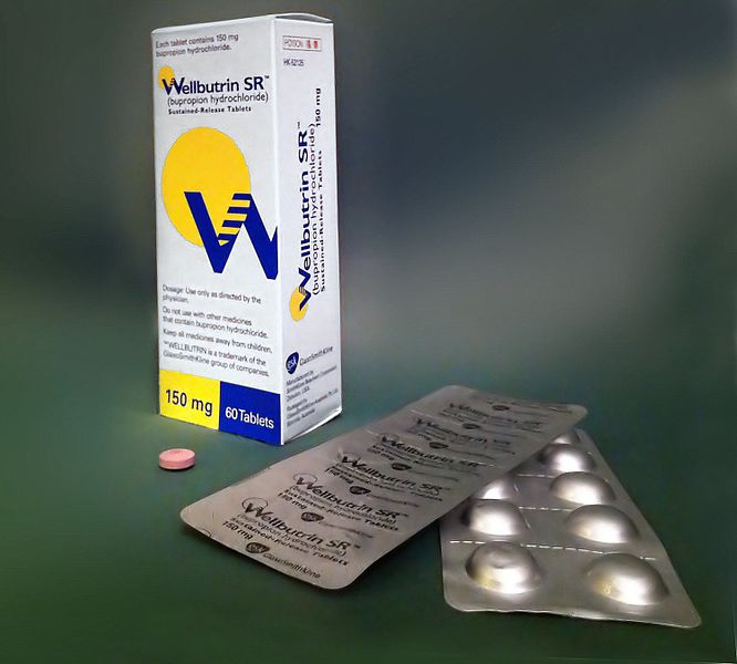
Image Credit: Сергей Фатеев, CC BY-SA 3.0, via Wikimedia Commons

Week 10 – EINSTEIN-PE

“Oral Rivaroxaban for the Treatment of Symptomatic Pulmonary Embolism”

by the EINSTEIN-PE Investigators

N Engl J Med. 2012 Apr 5;366(14):1287-97. [free full text]

Prior to the introduction of DOACs, the standard of care for treatment of acute VTE was treatment with a vitamin K antagonist (VKA, e.g. warfarin) bridged with LMWH. In 2010, the EINSTEIN-DVT study demonstrated the non-inferiority of rivaroxaban (Xarelto) versus VKA with an enoxaparin bridge in patients with acute DVT in the prevention of recurrent VTE. Subsequently, in this 2012 study, EINSTEIN-PE, the EINSTEIN investigators examined the potential role for rivaroxaban in the treatment of acute PE.

This open-label RCT compared treatment of acute PE (± DVT) with rivaroxaban (15mg PO BID x21 days, followed by 20mg PO daily) versus VKA with an enoxaparin 1mg/kg BID bridge until the INR was therapeutic for 2+ days and the patient had received at least 5 days of enoxaparin. Patients with cancer were not excluded if they had a life expectancy of ≥ 3 months, but they comprised only ~4.5% of the patient population. Treatment duration was determined by the discretion of the treating physician and was decided prior to randomization. Duration was also a stratifying factor in the randomization. The primary outcome was symptomatic recurrent VTE (fatal or nonfatal). The pre-specified noninferiority margin was 2.0 for the upper limit of the 95% confidence interval of the hazard ratio. The primary safety outcome was “clinically relevant bleeding.”

4833 patients were randomized. In the conventional-therapy group, the INR was in the therapeutic range 62.7% of the time. Symptomatic recurrent VTE occurred in 2.1% of patients in the rivaroxaban group and 1.8% of patients in the conventional-therapy group (HR 1.12, 95% CI 0.75–1.68, p = 0.003 for noninferiority). The p value for superiority of conventional therapy over rivaroxaban was 0.57. A first episode of “clinically relevant bleeding” occurred in 10.3% of the rivaroxaban group versus 11.4% of the conventional-therapy group (HR 0.90, 95% CI 0.76-1.07, p = 0.23).

In a large, open-label RCT, rivaroxaban was shown to be noninferior to standard therapy with a VKA + enoxaparin bridge in the treatment of acute PE. This was the first major RCT to demonstrate the safety and efficacy of a DOAC in the treatment of PE and led to FDA approval of rivaroxaban for the treatment of PE that same year. The following year, the AMPLIFY trial demonstrated that apixaban was noninferior to VKA + LMWH bridge in the prevention of recurrent VTE, and apixaban was also approved by the FDA for the treatment of PE. The 2016 Chest guidelines for Antithrombotic Therapy for VTE Disease recommend the DOACs rivaroxaban, apixaban, dabigatran, or edoxaban over VKA therapy in VTE not associated with cancer. In cancer-associated VTE, LMWH remains the recommended initial agent. (See the Week 1 – CLOT post.) As noted previously, a study in 2018 in NEJM demonstrated the noninferiority of edoxaban over LMWH in the treatment of cancer-associated VTE. Later that year, the SELECT-D trial compared rivaroxaban (Xarelto) to dalteparin and demonstrated a reduced rate of recurrence among patients treated with rivaroxaban (cumulative 6-month event rate of 4% versus 11%, HR 0.43, 95% CI 0.19–0.99) with no difference in rates of major bleeding but increased “clinically relevant nonmajor bleeding” within the rivaroxaban group.

Further Reading/References:
1. EINSTEIN-DVT @ NEJM
2. EINSTEIN-PE @ Wiki Journal Club
3. EINSTEIN-PE @ 2 Minute Medicine
4. AMPLIFY @ Wiki Journal Club
5. “Edoxaban for the Treatment of Cancer-Associated Venous Thromboembolism” NEJM 2018

Summary by Duncan F. Moore, MD

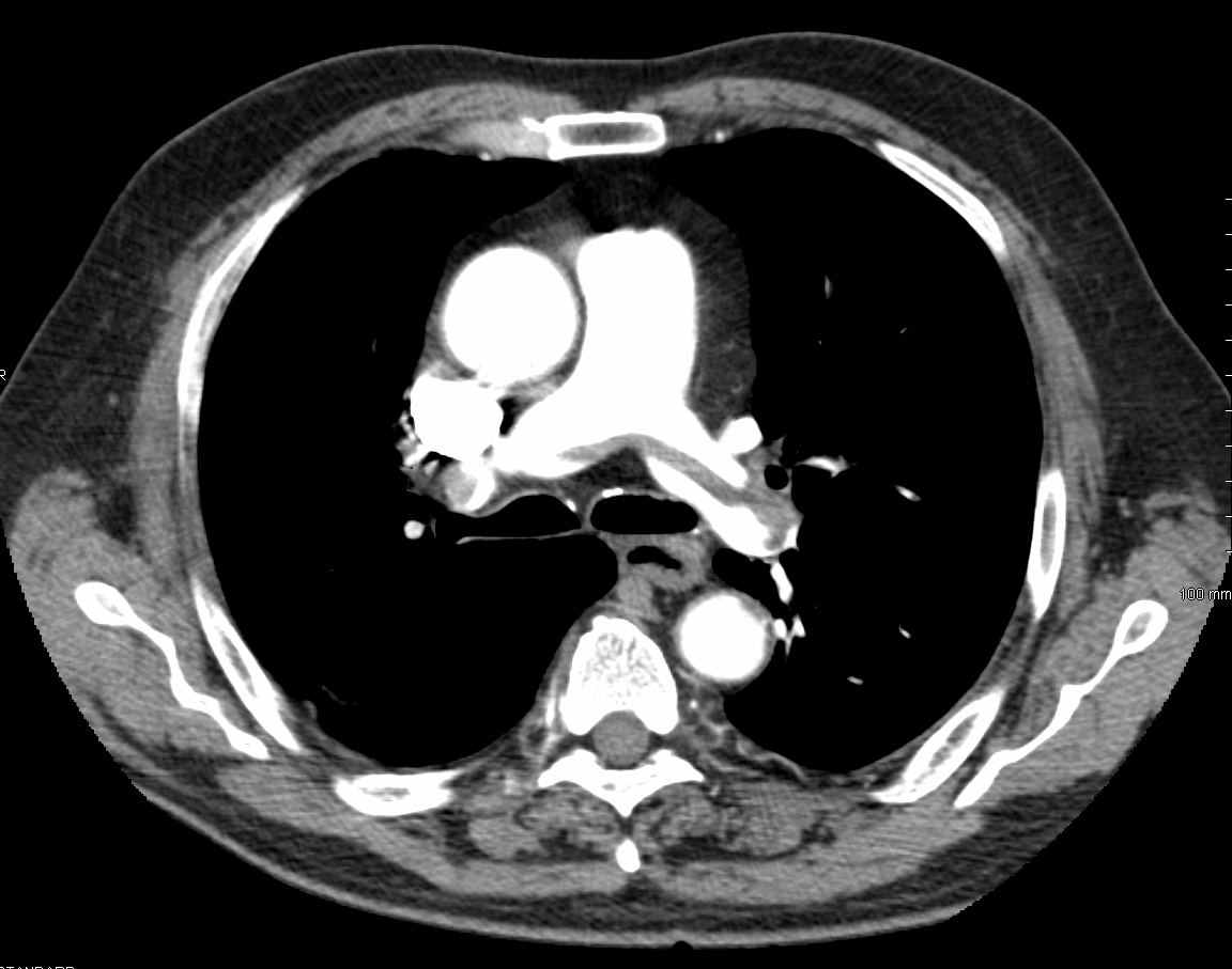
Image Credit: James Heilman, MD / CC BY-SA 4.0 / via WikiMedia Commons

Week 9 – Albumin in SBP

“Effect of Intravenous Albumin on Renal Impairment and Mortality in Patients with Cirrhosis and Spontaneous Bacterial Peritonitis”

N Engl J Med. 1999 Aug 5;341(6):403-9. [free full text]

Renal failure commonly develops in the setting of spontaneous bacterial peritonitis (SBP), and its development is a sensitive predictor of in-hospital mortality. The renal impairment is thought to stem from decreased effective arterial blood volume secondary to the systemic inflammatory response to the infection. In our current practice, there are certain circumstances in which we administer albumin early in the SBP disease course in order to reduce the risk of renal failure and mortality. Ultimately, our current protocol originated from the 1999 study of albumin in SBP by Sort et al.

The trial enrolled adults with SBP and randomized them to treatment with either cefotaxime and albumin infusion 1.5 gm/kg within 6hrs of enrollment, followed by 1 gm/kg on day 3 or cefotaxime alone. The primary outcome was the development of “renal impairment” (a “nonreversible” increase in BUN or Cr by more than 50% to a value greater than 30 mg/dL or 1.5 mg/dL, respectively) during hospitalization. The secondary outcome was in-hospital mortality.

126 patients were randomized. Both groups had similar baseline characteristics, and both had similar rates of resolution of infection. Renal impairment occurred in 10% of the albumin group and 33% of the cefotaxime-alone group (p = 0.02). In-hospital mortality was 10% in the albumin group and 29% in the cefotaxime-alone group (p = 0.01). 78% of patients that developed renal impairment died in-hospital, while only 3% of patients who did not develop renal impairment died. Plasma renin activity was significantly higher on days 3, 6, and 9 in the cefotaxime-alone group than in the albumin group, while there were no significant differences in MAP among the two groups at those time intervals. Multivariate analysis of all trial participants revealed that baseline serum bilirubin and creatinine were independent predictors of the development of renal impairment.

In conclusion, albumin administration reduces renal impairment and improves mortality in patients with SBP. The findings of this landmark trial were refined by a brief 2007 report by Sigal et al. entitled “Restricted use of albumin for spontaneous bacterial peritonitis.” “High-risk” patients, identified by baseline serum bilirubin of ≥ 4.0 mg/dL or Cr ≥ 1.0 mg/dL were given the intervention of albumin 1.5gm/kg on day 1 and 1gm/kg on day 3, and low-risk patients were not given albumin. None of the 15 low-risk patients developed renal impairment or died, whereas 12 of 21 (57%) of the high-risk group developed renal impairment, and 5 of the 21 (24%) died. The authors conclude that patients with bilirubin < 4.0 and Cr < 1.0 did not need scheduled albumin in the treatment of SBP. The current (2012) American Association for the Study of Liver Diseases guidelines for the management of adult patients with ascites due to cirrhosis do not definitively recommend criteria for albumin administration in SBP. Instead they summarize the aforementioned two studies. A 2013 meta-analysis of four reports/trials (including the two above) concluded that albumin infusion reduced renal impairment and improved mortality with pooled odds ratios approximately commensurate with those of the 1999 study by Sort et al. Ultimately, the current recommended practice per expert opinion is to perform albumin administration per the protocol outlined by Sigal et al. (2007).

References / Further Reading:
1. AASLD Guidelines for Management of Adult Patients with Ascites Due to Cirrhosis (skip to page 77)
2. Sigal et al. “Restricted use of albumin for spontaneous bacterial peritonitis”
3. Meta-analysis: “Albumin infusion improves outcomes of patients with spontaneous bacterial peritonitis: a meta-analysis of randomized trials”
4. Wiki Journal Club
5. 2 Minute Medicine

Summary by Duncan F. Moore, MD

Week 8 – 4S

“Randomised trial of cholesterol lowering in 4444 patients with coronary heart disease: the Scandinavian Simvastatin Survival Study (4S)”

Lancet. 1994 Nov 19;344(8934):1383-9 [free full text]

Statins are an integral part of modern primary and secondary prevention of atherosclerotic cardiovascular disease (ASCVD). Hypercholesterolemia is regarded as a major contributory factor to the development of atherosclerosis, and in the 1980s, a handful of clinical trials demonstrated reduction in MI/CAD incidence with cholesterol-lowering agents, such as cholestyramine and gemfibrozil. However, neither drug demonstrated a mortality benefit. By the late 1980s, there was much hope that the emerging drug class of HMG-CoA reductase inhibitors (statins) would confer a mortality benefit, given their previously demonstrated LDL-lowering effects. The 1994 Scandinavian Simvastatin Survival Study was the first large clinical trial to assess this hypothesis.

4444 adults ages 35-70 with a history of angina pectoris or MI and elevated serum total cholesterol (212 – 309 mg/dL) were recruited from 94 clinical centers in Scandinavia (and in Finland, which is technically a Nordic country but not a Scandinavian country…) and randomized to treatment with either simvastatin 20mg PO qPM or placebo. Dosage was increased at 12 weeks and 6 months to target a serum total cholesterol of 124 to 201 mg/dL. (Placebo patients were randomly uptitrated as well.) The primary endpoint was all-cause mortality. The secondary endpoint was time to first “major coronary event,” which included coronary deaths, nonfatal MI, resuscitated cardiac arrest, and definite silent MI per EKG.

The study was stopped early in 1994 after an interim analysis demonstrated a significant survival benefit in the treatment arm. At a mean 5.4 years of follow-up, 256 (12%) in the placebo group versus 182 (8%) in the simvastatin group had died (RR 0.70, 95% CI 0.58-0.85, p=0.0003, NNT = 30.1). The mortality benefit was driven exclusively by a reduction in coronary deaths. Dropout rates were similar (13% of placebo group and 10% of simvastatin group). The secondary endpoint, occurrence of a major coronary event, occurred in 622 (28%) of the placebo group and 431 (19%) of the simvastatin group (RR 0.66, 95% CI 0.59-0.75, p < 0.00001). Subgroup analyses of women and patients aged 60+ demonstrated similar findings for the primary and secondary outcomes. Over the entire course of the study, the average changes in lipid values from baseline in the simvastatin group were -25% total cholesterol, -35% LDL, +8% HDL, and -10% triglycerides. The corresponding percent changes from baseline in the placebo group were +1%, +1%, +1%, and +7%, respectively.

In conclusion, simvastatin therapy reduced mortality in patients with known CAD and hypercholesterolemia via reduction of major coronary events. This was a large, well-designed, double-blind RCT that ushered in the era of widespread statin use for secondary, and eventually, primary prevention of ASCVD. For further information about modern guidelines for the use of statins, please see the 2018 “ACC/AHA Multisociety Guideline on the Management of Blood Cholesterol” and the 2016 USPSTF guideline “Statin use for the Primary Prevention of Cardiovascular Disease in Adults: Preventive Medication”.

Finally, for history buffs interested in a brief history of the discovery and development of this drug class, please see this paper by Akira Endo.

References / Additional Reading:
1. 4S @ Wiki JournalClub
2. “2018 ACC/AHA Multisociety Guideline on the Management of Blood Cholesterol”
3. “Statin use for the Primary Prevention of Cardiovascular Disease in Adults: Preventive Medication” (2016)
4. UpToDate, “Society guideline links: Lipid disorders in adults”
5. “A historical perspective on the discovery of statins” (2010)

Summary by Duncan F. Moore, MD

Image Credit: Siol, CC BY-SA 3.0, via Wikimedia Commons

Week 7 – FUO

“Fever of Unexplained Origin: Report on 100 Cases”

Medicine (Baltimore). 1961 Feb;40:1-30. [free full text]

In our modern usage, fever of unknown origin (FUO) refers to a persistent unexplained fever despite an adequate medical workup. The most commonly used criteria for this diagnosis stem from this 1961 series by Petersdorf and Beeson.

This study analyzed a prospective cohort of patients evaluated at Yale’s hospital for FUO between 1952 and 1957. Their FUO criteria: 1) illness of more than three week’s duration, 2) fever higher than 101º F on several occasions, and 3) diagnosis uncertain after one week of study in hospital. After 126 cases had been noted, retrospective investigation was undertaken to determine the ultimate etiologies of the fevers. The authors winnowed this group to 100 cases based on availability of follow-up data and the exclusion of cases that “represented combinations of such common entities as urinary tract infection and thrombophlebitis.”

In 93 cases, “a reasonably certain diagnosis was eventually possible.” 6 of the 7 undiagnosed patients ultimately made a full recovery. Underlying etiologies (see table 1 on page 3) included: infectious 36% (with TB in 11%), neoplastic diseases 19%, collagen disease (e.g. SLE) 13%, pulmonary embolism 3%, benign non-specific pericarditis 2%, sarcoidosis 2%, hypersensitivity reaction 4%, cranial arteritis 2%, periodic disease 5%, miscellaneous disease 4%, factitious fever 3%, no diagnosis 7%.

Clearly, diagnostic modalities have improved markedly since this 1961 study. However, the core etiologies of infection, malignancy, and connective tissue disease/non-infectious inflammatory disease remain most prominent, while the percentage of patients with no ultimate diagnosis has been increasing (for example, see PMIDs 9413425, 12742800, and 17220753). Modifications to the 1961 criteria have been proposed (for example: 1 week duration of hospital stay not required if certain diagnostic measures have been performed) and implemented in recent FUO trials. One modern definition of FUO: fever ≥ 38.3º C, lasting at least 2-3 weeks, with no identified cause after three days of hospital evaluation or three outpatient visits. Per UpToDate, the following minimum diagnostic workup is recommended in suspected FUO: blood cultures, ESR or CRP, LDH, HIV, RF, heterophile antibody test, CK, ANA, TB testing, SPEP, and CT of abdomen and chest.

Further Reading/References:
1. “Fever of unknown origin (FUO). I A. prospective multicenter study of 167 patients with FUO, using fixed epidemiologic entry criteria. The Netherlands FUO Study Group.” Medicine (Baltimore). 1997 Nov;76(6):392-400.
2. “From prolonged febrile illness to fever of unknown origin: the challenge continues.” Arch Intern Med. 2003 May 12;163(9):1033-41.
3. “A prospective multicenter study on fever of unknown origin: the yield of a structured diagnostic protocol.” Medicine (Baltimore). 2007 Jan;86(1):26-38.
4. UpToDate, “Approach to the Adult with Fever of Unknown Origin”
5. “Robert Petersdorf, 80, Major Force in U.S. Medicine, Dies” The New York Times, 2006

Summary by Duncan F. Moore, MD

Week 6 – Bicarbonate and Progression of CKD

“Bicarbonate Supplementation Slows Progression of CKD and Improves Nutritional Status”

J Am Soc Nephrol. 2009 Sep;20(9):2075-84. [free full text]

Metabolic acidosis is a common complication of advanced CKD. Some animal models of CKD have suggested that worsening metabolic acidosis is associated with worsening proteinuria, tubulointerstitial fibrosis, and acceleration of decline of renal function. Short-term human studies have demonstrated that bicarbonate administration reduces protein catabolism and that metabolic acidosis is an independent risk factor for acceleration of decline of renal function. However, until this 2009 study by de Brito-Ashurst et al., there were no long-term studies demonstrating the beneficial effects of oral bicarbonate administration on CKD progression and nutritional status.

The study enrolled CKD patients with CrCl 15-30ml/min and plasma bicarbonate 16-20 mEq/L and randomized them to treatment with either sodium bicarbonate 600mg PO TID (with protocolized uptitration to achieve plasma HCO3  ≥ 23 mEq/L) for 2 years, or to routine care. The primary outcomes were: 1) the decline in CrCl at 2 years, 2) “rapid progression of renal failure” (defined as decline of CrCl > 3 ml/min per year), and 3) development of ESRD requiring dialysis. Secondary outcomes included 1) change in dietary protein intake, 2) change in normalized protein nitrogen appearance (nPNA), 3) change in serum albumin, and 4) change in mid-arm muscle circumference.

134 patients were randomized, and baseline characteristics were similar among the two groups. Serum bicarbonate levels increased significantly in the treatment arm. (See Figure 2.) At two years, CrCl decline was 1.88 ml/min in the treatment group vs. 5.93 ml/min in the control group (p < 0.01). Rapid progression of renal failure was noted in 9% of intervention group vs. 45% of the control group (RR 0.15, 95% CI 0.06–0.40, p < 0.0001, NNT = 2.8), and ESRD developed in 6.5% of the intervention group vs. 33% of the control group (RR 0.13, 95% CI 0.04–0.40, p < 0.001; NNT = 3.8). Regarding nutritional status, dietary protein intake increased in the treatment group relative to the control group (p < 0.007). Normalized protein nitrogen appearance decreased in the treatment group and increased in the control group (p < 0.002). Serum albumin increased in the treatment group but was unchanged in the control group, and mean mid-arm muscle circumference increased by 1.5 cm in the intervention group vs. no change in the control group (p < 0.03).

In conclusion, oral bicarbonate supplementation in CKD patients with metabolic acidosis reduces the rate of CrCl decline and progression to ESRD and improves nutritional status. Primarily on the basis of this study, the KDIGO 2012 guidelines for the management of CKD recommend oral bicarbonate supplementation to maintain serum bicarbonate within the normal range (23-29 mEq/L). This is a remarkably cheap and effective intervention. Importantly, the rates of adverse events, particularly worsening hypertension and increasing edema, were unchanged among the two groups. Of note, sodium bicarbonate induces much less volume expansion than a comparable sodium load of sodium chloride.

In their discussion, the authors suggest that their results support the hypothesis of Nath et al. (1985) that “compensatory changes [in the setting of metabolic acidosis] such as increased ammonia production and the resultant complement cascade activation in remnant tubules in the declining renal mass [are] injurious to the tubulointerstitium.” The hypercatabolic state of advanced CKD appears to be mitigated by bicarbonate supplementation. The authors note that “an optimum nutritional status has positive implications on the clinical outcomes of dialysis patients, whereas [protein-energy wasting] is associated with increased morbidity and mortality.”

Limitations to this trial include its open-label, no-placebo design. Also, the applicable population is limited by study exclusion criteria of morbid obesity, overt CHF, and uncontrolled HTN.

Further Reading:
1. Nath et al. “Pathophysiology of chronic tubulo-interstitial disease in rats: Interactions of dietary acid load, ammonia, and complement component-C3” (1985)
2. KDIGO 2012 Clinical Practice Guideline for the Evaluation and Management of Chronic Kidney Disease (see page 89)
3. UpToDate, “Pathogenesis, consequences, and treatment of metabolic acidosis in chronic kidney disease”

Summary by Duncan F. Moore, MD

Week 5 – Dexamethasone in Bacterial Meningitis

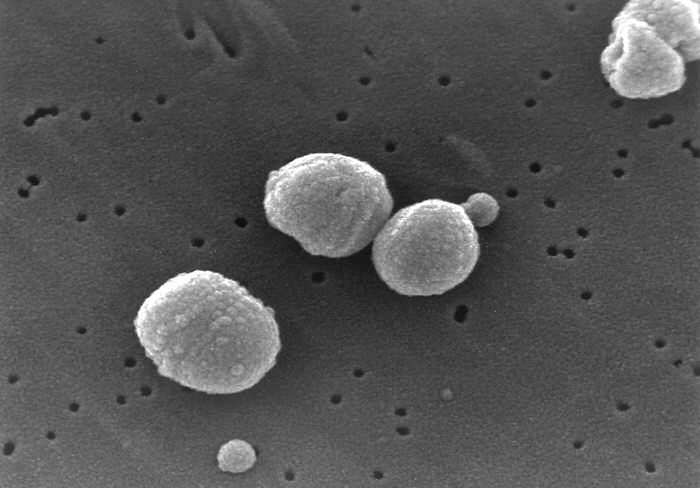
Streptococcus pneumoniae

“Dexamethasone in Adults With Bacterial Meningitis”

N Engl J Med 2002; 347:1549-1556 [free full text]

The current standard of care in the treatment of suspected bacterial meningitis in the developed world includes the administration of dexamethasone prior to or at the time of antibiotic initiation. The initial evaluation of this practice in part stemmed from animal studies, which demonstrated that dexamethasone reduced CSF concentrations of inflammatory markers as well as neurologic sequelae after meningitis. RCTs in the pediatric literature also demonstrated clinical benefit. The best prospective trial in adults was this 2002 study by de Gans et al.

The trial enrolled adults with suspected meningitis and randomized them to either dexamethasone 10mg IV q6hrs x4 days started 15-20 minutes before the first IV antibiotics or a placebo IV with the same administration schedule. The primary outcome was the Glasgow Outcome Scale at 8 weeks (1 = death, 2 = vegetative state, 3 = unable to live independently, 4 = unable to return to school/work, 5 = able to return to school/work). Secondary outcomes included death and focal neurologic abnormalities. Subgroup analyses were performed by organism.

301 patients were randomized. At 8 weeks, 15% of dexamethasone patients compared with 25% of placebo patients had an unfavorable outcome of Glasgow Outcome Scale score 1-4 (RR 0.59, 95% CI 0.37 – 0.94, p= 0.03). Among patients with pneumococcal meningitis, 26% of dexamethasone patients compared with 52% of placebo patients had an unfavorable outcome. There was no significant difference among treatment arms within the subgroup of patients infected with meningococcal meningitis. Overall, death occurred in 7% of dexamethasone patients and 15% of placebo patients (RR 0.48, 95% CI 0.24 – 0.96, p = 0.04). In pneumococcal meningitis, 14% of dexamethasone patients died, and 34% of placebo patients died.  There was no difference in rates of focal neurologic abnormalities or hearing loss in either treatment arm (including within any subgroup).

In conclusion, early adjunctive dexamethasone improves mortality in bacterial meningitis. As noted in the above subgroup analysis, this benefit appears to be driven by the efficacy within the pneumococcal meningitis subgroup. Of note, the standard initial treatment regimen in this study was amoxicillin 2gm q4hrs for 7-10 days rather than our standard ceftriaxone + vancomycin +/- ampicillin. Largely on the basis of this study alone, the IDSA guidelines for the treatment of bacterial meningitis (2004) recommend dexamethasone 0.15 mg/kg q6hrs for 2-4 days with first dose administered 10-20 min before or concomitant with initiation of antibiotics. Dexamethasone should be continued only if CSF Gram stain, CSF culture, or blood cultures are consistent with pneumococcus.

References / Further Reading:
1. IDSA guidelines for management of bacterial meningitis (2004)
2. Wiki Journal Club
3. 2 Minute Medicine

Summary by Duncan F. Moore, MD

Image Credit: CDC / Dr. Richard Facklam, US Public Domain, via Public Health Image Library

Week 4 – ARDSNet

“Ventilation with Lower Tidal Volumes as Compared with Traditional Tidal Volumes for Acute Lung Injury and the Acute Respiratory Distress Syndrome”

by the Acute Respiratory Distress Syndrome Network (ARDSNet)

N Engl J Med. 2000 May 4;342(18):1301-8. [free full text]

Acute respiratory distress syndrome (ARDS) is an inflammatory and highly morbid lung injury found in many critically ill patients. In the 1990s, it was hypothesized that overdistention of aerated lung volumes and elevated airway pressures might contribute to the severity of ARDS, and indeed some work in animal models supported this theory. Prior to the ARDSNet study, four randomized trials had been conducted to investigate the possible protective effect of ventilation with lower tidal volumes, but their results were conflicting.

The ARDSNet study enrolled patients with ARDS (diagnosed within 36 hours) to either a lower initial tidal volume of 6ml/kg, downtitrated as necessary to maintain plateau pressure ≤ 30 cm H2O, or to the “traditional” therapy of an initial tidal volume of 12 ml/kg, downtitrated as necessary to maintain plateau pressure ≤ 50 cm of water. The primary outcomes were in-hospital mortality and ventilator-free days within the first 28 days. Secondary outcomes included number of days without organ failure, occurrence of barotrauma, and reduction in IL-6 concentration from day 0 to day 3.

861 patients were randomized before the trial was stopped early due to the increased mortality in the control arm noted during interim analysis. In-hospital mortality was 31.0% in the lower tidal volume group and 39.8% in the traditional tidal volume group (p = 0.007, NNT = 11.4). Ventilator free days were 12±11 in the lower tidal volume group vs. 10±11 in the traditional group (n = 0.007). The lower tidal volume group had more days without organ failure (15±11 vs. 12±11, p = 0.006). There was no difference in rates of barotrauma among the two groups. Decrease in IL-6 concentration between days 0 and 3 was greater in the low tidal volume group (p < 0.001), and IL-6 concentration at day 3 was lower in the low tidal volume group (p = 0.002).

In summary, low tidal volume ventilation decreases mortality in ARDS relative to “traditional” tidal volumes. The authors felt that this study confirmed the results of prior animal models and conclusively answered the question of whether or not low tidal volume ventilation provided a mortality benefit. In fact, in the years following, low tidal volume ventilation became the standard of care, and a robust body of literature followed this study to further delineate a “lung-protective strategy.” Critics of the study noted that, at the time of the study, the “traditional” (standard of care) tidal volume in ARDS was less than the 12 ml/kg used in the comparison arm. (Non-enrolled patients at the participating centers were receiving a mean tidal volume of 10.3 ml/kg.) Thus not only was the trial making a comparison to a faulty control, but it was also potentially harming patients in the control arm. An excellent summary of the ethical issues and debate regarding this specific issue and regarding control arms of RCTs in general can be found here.

Corresponding practice point from Dr. Sonti and Dr. Vinayak and their Georgetown Critical Care Top 40: “Low tidal volume ventilation is the standard of care in patients with ARDS (P/F < 300). Use ≤ 6 ml/kg predicted body weight, follow plateau pressures, and be cautious of mixed modes in which you set a tidal volume but the ventilator can adjust and choose a larger one.”

PulmCCM is an excellent blog, and they have a nice page reviewing this topic and summarizing some of the research and guidelines that have followed.

Further Reading/References:
1. ARDSNet @ Wiki Journal Club
2. ARDSNet @ 2 Minute Medicine
3. PulmCCM “Mechanical Ventilation in ARDS: Research Update”
4. Georgetown Critical Care Top 40, page 6
5. PulmCCM “In ARDS, substandard ventilator care is the norm, not the exception.” 2017.

Summary by Duncan F. Moore, MD

Week 3 – NICE-SUGAR

“Intensive versus Conventional Glucose Control in Critically Ill Patients”

by the Normoglycemia in Intensive Care Evaluation–Survival Using Glucose Algorithm Regulation (NICE-SUGAR) investigators

N Engl J Med 2009;360:1283-97. [free full text]

On the wards we often hear 180 mg/dL used as the upper limit of acceptable for blood glucose with the understanding that tighter glucose control in inpatients can lead to more harm than benefit. The relevant evidence base comes from ICU populations, with scant direct data in non-ICU patients. The 2009 NICE-SUGAR study is the largest and best among this evidence base.

The study randomized ICU patients (expected to require 3 or more days of ICU-level care) to either “intensive” glucose control (target glucose 81 to 108 mg/dL) or conventional glucose control (target of less than 180 mg/dL). The primary outcome was 90-day all-cause mortality.

6104 patients were randomized to the two arms, and both groups had similar baseline characteristics. 27.5% of patients in the intensive-control group died versus 24.9% in the conventional-control group (OR 1.14, 95% CI 1.02-1.28, p= 0.02). Severe hypoglycemia (< 40 mg/dL) was found in 6.8% of intensive patients but only 0.5% of conventional patients.

In conclusion, intensive glucose control increases mortality in ICU patients. The fact that only 20% of these patients had diabetes mellitus suggests that much of the hyperglycemia treated in this study (97% of intensive group received insulin, 69% of conventional) was from stress, critical illness, and corticosteroid use. For ICU patients, intensive insulin therapy is clearly harmful, but the ideal target glucose range remains controversial and by expert opinion appears to be 140-180. For non-ICU inpatients with or without diabetes mellitus, the ideal glucose target is also unclear – the ADA recommends 140-180, and the Endocrine Society recommends a pre-meal target of < 140 and random levels < 180.

References / Further Reading:
1. ADA Standards of Medical Care in Diabetes 2016 (skip to page S99)
2. NICE-SUGAR @ Wiki Journal Club

Summary by Duncan F. Moore, MD