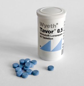
Week 52 – Symptom-Triggered Benzodiazepines in Alcohol Withdrawal

“Symptom-Triggered vs Fixed-Schedule Doses of Benzodiazepine for Alcohol Withdrawal”

Arch Intern Med. 2002 May 27;162(10):1117-21. [free full text]

Treatment of alcohol withdrawal with benzodiazepines has been the standard of care for decades. However, in the 1990s, benzodiazepine therapy for alcohol withdrawal was generally given via fixed doses. In 1994, a double-blind RCT by Saitz et al. demonstrated that symptom-triggered therapy based on responses to the CIWA-Ar scale reduced treatment duration and the amount of benzodiazepine used relative to a fixed-schedule regimen. This trial had little immediate impact in the treatment of alcohol withdrawal. The authors of the 2002 double-blind RCT sought to confirm the findings from 1994 in a larger population that did not exclude patients with a history of seizures or severe alcohol withdrawal.

The trial enrolled consecutive patients admitted to the inpatient alcohol treatment units at two European universities (excluding those with “major cognitive, psychiatric, or medical comorbidity”) and randomized them to treatment with either scheduled placebo (30mg q6hrs x4, followed by 15mg q6hrs x8) with additional PRN oxazepam 15mg for CIWA score 8-15 and 30mg for CIWA score > 15 or to treatment with scheduled oxazepam (30mg q6hrs x4, followed by 15mg q6hrs x8) with additional PRN oxazepam 15mg for CIWA score 8-15 and 30mg for CIWA score > 15.

The primary outcomes were cumulative oxazepam dose at 72 hours and duration of treatment with oxazepam. Subgroup analysis included the exclusion of symptomatic patients who did not require any oxazepam. Secondary outcomes included incidence of seizures, hallucinations, and delirium tremens at 72 hours.

Results:
117 patients completed the trial. 56 had been randomized to the symptom-triggered group, and 61 had been randomized to the fixed-schedule group. The groups were similar in all baseline characteristics except that the fixed-schedule group had on average a 5-hour longer interval since last drink prior to admission. While only 39% of the symptom-triggered group actually received oxazepam, 100% of the fixed-schedule group did (p < 0.001). Patients in the symptom-triggered group received a mean cumulative dose of 37.5mg versus 231.4mg in the fixed-schedule group (p < 0.001). The mean duration of oxazepam treatment was 20.0 hours in the symptom-triggered group versus 62.7 hours in the fixed-schedule group. The group difference in total oxazepam dose persisted even when patients who did not receive any oxazepam were excluded. Among patients who did receive oxazepam, patients in the symptom-triggered group received 95.4 ± 107.7mg versus 231.4 ± 29.4mg in the fixed-dose group (p < 0.001). Only one patient in the symptom-triggered group sustained a seizure. There were no seizures, hallucinations, or episodes of delirium tremens in any of the other 116 patients. The two treatment groups had similar quality-of-life and symptom scores aside from slightly higher physical functioning in the symptom-triggered group (p < 0.01). See Table 2.

Implication/Discussion:
Symptom-triggered administration of benzodiazepines in alcohol withdrawal led to a six-fold reduction in cumulative benzodiazepine use and a much shorter duration of pharmacotherapy than fixed-schedule administration. This more restrictive and responsive strategy did not increase the risk of major adverse outcomes such as seizure or DTs and also did not result in increased patient discomfort.

Overall, this study confirmed the findings of the landmark study by Saitz et al. from eight years prior. Additionally, this trial was larger and did not exclude patients with a prior history of withdrawal seizures or severe withdrawal. The fact that both studies took place in inpatient specialty psychiatry units limits their generalizability to our inpatient general medicine populations.

Why the initial 1994 study did not gain clinical traction remains unclear. Both studies have been well-cited over the ensuing decades, and the paradigm has shifted firmly toward symptom-triggered benzodiazepine regimens using the CIWA scale. While a 2010 Cochrane review cites only the 1994 study, Wiki Journal Club and 2 Minute Medicine have entries on this 2002 study but not on the equally impressive 1994 study.

Further Reading/References:
1. “Individualized treatment for alcohol withdrawal. A randomized double-blind controlled trial.” JAMA. 1994.
2. Clinical Institute Withdrawal Assessment of Alcohol Scale, Revised (CIWA-Ar)
3. Wiki Journal Club
4. 2 Minute Medicine
5. “Benzodiazepines for alcohol withdrawal.” Cochrane Database Syst Rev. 2010

Summary by Duncan F. Moore, MD

Image Credit: VisualBeo, CC BY-SA 3.0, via Wikimedia Commons

Week 51 – LOTT

“A Randomized Trial of Long-Term Oxygen for COPD with Moderate Desaturation”

by the Long-Term Oxygen Treatment Trial (LOTT) Research Group

N Engl J Med. 2016 Oct 27;375(17):1617-1627. [free full text]

The long-term treatment of severe resting hypoxemia (SpO2 < 89%) in COPD with supplemental oxygen has been a cornerstone of modern outpatient COPD management since its mortality benefit was demonstrated circa 1980. Subsequently, the utility of supplemental oxygen in COPD patients with moderate resting daytime hypoxemia (SpO2 89-93%) was investigated in trials in the 1990s; however, such trials were underpowered to assess mortality benefit. Ultimately, the LOTT trial was funded by the NIH and Centers for Medicare and Medicaid Services (CMS) primarily to determine if there was a mortality benefit to supplemental oxygen in COPD patients with moderate hypoxemia as well to analyze as numerous other secondary outcomes, such as hospitalization rates and exercise performance.

The LOTT trial was originally planned to enroll 3500 patients. However, after 7 months the trial had randomized only 34 patients, and mortality had been lower than anticipated. Thus in late 2009 the trial was redesigned to include broader inclusion criteria (now patients with exercise-induced hypoxemia could qualify) and the primary endpoint was broadened from mortality to a composite of time to first hospitalization or death.

The revised LOTT trial enrolled COPD patients with moderate resting hypoxemia (SpO2 89-93%) or moderate exercise-induced desaturation during the 6-minute walk test (SpO2 ≥ 80% for ≥ 5 minutes and < 90% for ≥ 10 seconds). Patients were randomized to either supplemental oxygen (24-hour oxygen if resting SpO2 89-93%, otherwise oxygen only during sleep and exercise if the desaturation occurred only during exercise) or to usual care without supplemental oxygen. Supplemental oxygen flow rate was 2 liters per minute and could be uptitrated by protocol among patients with exercise-induced hypoxemia. The primary outcome was time to composite of first hospitalization or death. Secondary outcomes included hospitalization rates, lung function, performance on 6-minute walk test, and quality of life.

368 patients were randomized to the supplemental-oxygen group and 370 to the no-supplemental-oxygen group. Of the supplemental-oxygen group, 220 patients were prescribed 24-hour oxygen support, and 148 were prescribed oxygen for use during exercise and sleep only. Median duration of follow-up was 18.4 months. Regarding the primary outcome, there was no group difference in time to death or first hospitalization (p = 0.52 by log-rank test). See Figure 1A. Furthermore, there were no treatment-group differences in the primary outcome among patients of the following pre-specified subgroups: type of oxygen prescription, “desaturation profile,” race, sex, smoking status, SpO2 nadir during 6-minute walk, FEV1, BODE  index, SF-36 physical-component score, BMI, or history of anemia. Patients with a COPD exacerbation in the 1-2 months prior to enrollment, age 71+ at enrollment, and those with lower Quality of Well-Being Scale score at enrollment all demonstrated benefit from supplemental O2, but none of these subgroup treatment effects were sustained when the analyses were adjusted for multiple comparisons. Regarding secondary outcomes, there were no treatment-group differences in rates of all-cause hospitalizations, COPD-related hospitalizations, or non-COPD-related hospitalizations, and there were no differences in change from baseline measures of quality of life, anxiety, depression, lung function, and distance achieved in 6-minute walk.

The LOTT trial presents compelling evidence that there is no significant benefit, mortality or otherwise, of oxygen supplementation in patients with COPD and either moderate hypoxemia at rest (SpO2 > 88%) or exercise-induced hypoxemia. Although this trial’s substantial redesign in its early course is noted, the trial still is our best evidence to date about the benefit (or lack thereof) of oxygen in this patient group. As acknowledged by the authors, the trial may have had significant selection bias in referral. (Many physicians did not refer specific patients for enrollment because “they were too ill or [were believed to have benefited] from oxygen.”) Another notable limitation of this study is that nocturnal oxygen saturation was not evaluated. The authors do note that “some patients with COPD and severe nocturnal desaturation might benefit from nocturnal oxygen supplementation.”

For further contemporary contextualization of the study, please see the excellent post at PulmCCM from 11/2016. Included in that post is a link to an overview and Q&A from the NIH regarding the LOTT study.

References / Additional Reading:
1. PulmCCM, “Long-term oxygen brought no benefits for moderate hypoxemia in COPD”
2. LOTT @ 2 Minute Medicine
3. LOTT @ ClinicalTrials.gov
4. McDonald, J.H. 2014. Handbook of Biological Statistics (3rd ed.). Sparky House Publishing, Baltimore, Maryland.
5. Centers for Medicare and Medicaid Services, “Certificate of Medical Necessity CMS-484– Oxygen”
6. Ann Am Thorac Soc. 2018 Dec;15(12):1369-1381. “Optimizing Home Oxygen Therapy. An Official American Thoracic Society Workshop Report.”

Summary by Duncan F. Moore, MD

Image Credit: Patrick McAleer, CC BY-SA 2.0 UK, via Wikimedia Commons

Week 50 – PARADIGM-HF

“Angiotensin-Neprilysin Inhibition versus Enalapril in Heart Failure”

N Engl J Med. 2014 Sep 11;371(11):993-1004. [free full text]

Background:
Thanks to the CONSENSUS and SOLVD trials, angiotensin-converting enzyme (ACE) inhibitors have been a cornerstone of the treatment of heart failure with reduced ejection fraction (HFrEF) for years. Neprilysin is a neutral endopeptidase that degrades several peptides, including natriuretic peptides, bradykinin, and adrenomedullin. Inhibiting neprilysin increases levels of these substances and thus counteracts the neurohormonal overactivation of heart failure (which would otherwise lead to vasoconstriction, sodium retention, and maladaptive remodeling). Prior experimental data has demonstrated that, in terms of cardiovascular outcomes, neprilysin inhibition with an ARB is superior to ARB monotherapy. However, a clinical trial of concurrent neprilysin-inhibitor and ACE inhibitor therapy resulted in unacceptably high rates of serious angioedema. This study sought to show improved cardiac and mortality outcomes with neprilysin inhibition plus an ARB when compared to enalapril alone.

The study enrolled adults with NYHA class II, III, or IV heart failure, LVEF ≤ 35%, and BNP ≥ 150 or NT-proBNP ≥ 600. Pertinent exclusion criteria included symptomatic hypotension, SBP < 100mmHg at screening or 95mmHg at randomization, eGFR < 30 or decrease in eGFR by 25% between screening and randomization, K+ > 5.2, or history of angioedema/side effects to ACE inhibition or ARBs. Patients were randomized to treatment with either sacubitril/valsartan 200mg BID or to enalapril 10mg BID. (Screened patients were initially given sacubitril/valsartan followed by enalapril in single blinded run-in phases in order to ensure similar tolerance of the drugs prior to randomization.) The primary outcome was a composite of death from cardiovascular causes or first hospitalization for heart failure. Selected secondary outcomes included: 1) change from baseline in the clinical summary score of the Kansas City Cardiomyopathy Questionnaire (KCCQ), 2) time to new-onset atrial fibrillation, and 3) time to first occurrence of decline in renal function.

Results:
4187 patients were randomized to the sacubitril/valsartan group, and 4212 were randomized to the enalapril group.

The primary endpoint (composite death due to cardiovascular causes or first hospitalization for HF) occurred in 914 patients (21.8%) in the sacubitril/valsartan group and 1117 patients (26.5%) in the enalapril group (p < 0.001; NNT = 21). Death due to cardiovascular causes occurred 558 times in the sacubitril/valsartan group and 693 times in the enalapril group (13.3% vs. 16.5%, p < 0.001; NNT = 31). Hospitalization for heart failure occurred (at least once) 537 times in the sacubitril/valsartan group and 658 times in the enalapril group (12.8% vs. 15.6%, p < 0.001; NNT = 36).

Regarding secondary outcomes, the mean change in KCCQ score was a reduction of 2.99 points (i.e. a worsening of symptoms) in the sacubitril/valsartan group versus a reduction of 4.63 points in the enalapril group (p = 0.001). There was no significant group difference in time to new-onset atrial fibrillation or time to diminished renal function.

Regarding safety outcomes, patients in the sacubitril/valsartan group were more likely to have symptomatic hypotension compared to patients in the enalapril group (14.0% vs. 9.2%; p < 0.001; NNH = 21). However, patients in the enalapril group were more likely to have cough, serum creatinine ≥ 2.5, or potassium ≥6.0 compared to sacubitril/valsartan (p value varies, all significant). There was no group difference in rates of angioedema (p = 0.13).

Implication/Discussion:
In patients with HFrEF, inhibition of both angiotensin II and neprilysin with sacubitril/valsartan significantly reduced the risk of cardiovascular death or hospitalization for heart failure when compared to treatment with enalapril alone.

This study had several strengths. The treatment with sacubitril/valsartan was compared to treatment with a dose of enalapril that had previously been shown to reduce mortality when compared with placebo. Furthermore, the study used a run-in phase to ensure that patients could tolerate an enalapril dose that had previously been shown to reduce mortality. Finally, more patients in the enalapril group than in the sacubitril/valsartan group stopped the study drug due to adverse effects (12.3% vs. 10.7%, p = 0.03).

This study ushered in a new era in heart failure management and added a new medication class – Angiotensin Receptor-Neprilysin Inhibitors or ARNIs – to the arsenal of available heart failure drugs. Entresto (sacubitril/valsartan), the ARNI posterchild, has been advertised widely over the past several years. However, clinical use so far has been lower than expected (see here).

The 2017 ACC/AHA update to the guidelines for management of symptomatic HFrEF states that primary inhibition of the renin-angiotensin system with an ARNI in conjunction with evidence-based beta blockade and aldosterone antagonism is a Class I recommendation (Level B evidence). However, it does not favor this regimen over the Level-A-evidence regimens of an ARB or ACE inhibitor substituted for the ARNI. Yet the new guidelines also state that patients who have chronic symptomatic HFrEF of NYHA class II or III and tolerate an ACE inhibitor or ARB should substitute an ARNI for the ACE inhibitor or ARB in order to further reduce morbidity and mortality (Class I recommendation, level B evidence). See pages 15 and 17 here to read the details.

Bottom line: Among patients with symptomatic HFrEF, treatment with an ARNI reduces cardiovascular mortality and HF hospitalizations when compared to treatment with enalapril. Due to this study’s impact, the use of ARNIs is now a Class I recommendation by the 2017 ACC/AHA guidelines for the treatment of HFrEF. Despite its higher cost, the use of sacubitril/valsartan appears to be cost-effective in terms of QALYs gained.

Further Reading/References:
1. Wiki Journal Club
2. 2 Minute Medicine
3. ACC/AHA 2017 Focused Update for Guideline Management of Heart Failure.
4. CardioBrief, “After Slow Start Entresto Is Poised For Takeoff.”
5. PARAGON-HF @ ClinicalTrials.gov
6. McMurray et al., “Cost-effectiveness of sacubitril/valsartan in the treatment of heart failure with reduced ejection fraction.” Heart, 2017.

Summary by Patrick Miller, MD

Week 49 – Donor-Feces Infusion for Recurrent C. difficile

“Duodenal Infusion of Donor Feces for Recurrent Clostridium difficile

N Engl J Med. 2013 Jan 31;368(5):407-15. [free full text]

Clostridioides (formerly Clostridium) difficile infection (CDI) is a common, increasingly prevalent, and increasingly recurrent disease. As discussed in the 2017-2018 Academic Year Week 43 post, the IDSA/SHEA guidelines published March 2018 now list vancomycin PO as first line treatment for initial, non-severe CDI. These guidelines also list fecal microbiota transplantation (FMT) as an option for treatment of a second or subsequent recurrence of CDI. FMT received a rating of “Strong [recommendation] / Moderate [level of evidence]” for this indication thanks to this 2013 trial by van Nood et al. – the first prospective RCT to compare antibiotic therapy to FMT in recurrent CDI.

This single-academic-center (Netherlands), open-label, randomized controlled trial compared three regimens for the treatment of recurrent CDI. One treatment arm received vancomycin 500mg PO QID x4-5 days followed by bowel lavage and then infusion of donor feces through nasoduodenal tube, another treatment arm received a standard 14-day vancomycin 500mg PO QID regimen, and the final treatment arm received a standard 14-day vancomycin regimen with additional bowel lavage on day 4 or 5. The primary endpoint was cure without relapse by 10 weeks.

43 patients were randomized prior to the termination of the trial due to the markedly higher rates of recurrent CDI among patients who did not receive FMT. Regarding the primary outcome, 13 (81%) of the FMT group were cured after the first infusion (and remained so) at 10 weeks, whereas resolution of CDI occurred in only 4 (31%) of the vancomycin-alone group and in only 3 (23%) of the vancomycin + bowel lavage group (p < 0.001 for both pairwise comparisons vs. FMT).

In this randomized controlled trial, fecal microbiota transplantation was superior to both vancomycin and vancomycin plus bowel lavage in the cure of recurrent Clostridioides difficile infection. Although this trial was small, its effect was enormous. As mentioned above, FMT is now recommended by guidelines for the treatment of multiply-recurrent CDI. FMT has been the subject of numerous published and ongoing trials, including this notable 2017 study by Kao et al. that demonstrated noninferiority of FMT delivered via oral capsules versus “conventional” colonoscopic delivery.

Further Reading/References:
1. Wiki Journal Club
2. 2 Minute Medicine
3. 2017 Update to IDSA/SHEA Clinical Practice Guidelines for Clostridium difficile Infection
4. Kao et. al, “Effect of Oral Capsule- vs Colonoscopy-Delivered Fecal Microbiota Transplantation on Recurrent Clostridium difficile Infection.” JAMA. 2017;318(20):1985-1993.
5. IDSA, “Fecal Microbiota Transplantation”
6. Food and Drug Administration, “Enforcement Policy Regarding Investigational New Drug Requirements for Use of Fecal Microbiota for Transplantation to Treat Clostridium difficile Infection Not Responsive to Standard Therapies”

Summary by Duncan F. Moore, MD

Image Credit: CDC/ Lois S. Wiggs (PHIL #6260), Public Domain, via Wikimedia Commons

Week 48 – Sepsis-3

“The Third International Consensus Definitions for Sepsis and Septic Shock (Sepsis-3)”

JAMA. 2016 Feb 23;315(8):801-10. [free full text]

In practice, we recognize sepsis as a potentially life-threatening condition that arises secondary to infection. Because the SIRS criteria were of limited sensitivity and specificity in identifying sepsis and because our understanding of the pathophysiology of sepsis had purportedly advanced significantly during the interval since the last sepsis definition, an international task force of 19 experts was convened to define and prognosticate sepsis more effectively. The resulting 2016 Sepsis-3 definition was the subject of immediate and sustained controversy.

In the words of Sepsis-3, sepsis simply “is defined as life-threatening organ dysfunction caused by a dysregulated host response to infection.” The paper further defines organ dysfunction in terms of a threshold change in the SOFA score by 2+ points. However, the authors state that “the SOFA score is not intended to be used as a tool for patient management but as a means to clinically characterize a septic patient.” The authors note that qSOFA, an easier tool introduced in this paper, can identify promptly at the bedside patients “with suspected infection who are likely to have a prolonged ICU stay or die in the hospital.” A positive screen on qSOFA is identified as 2+ of the following: AMS, SBP ≤ 100, or respiratory rate ≥ 22. At the time of this endorsement of qSOFA, the tool had not been validated prospectively. Finally, septic shock was defined as sepsis with persistent hypotension requiring vasopressors to maintain MAP ≥ 65 and with a serum lactate > 2 despite adequate volume resuscitation.

As noted contemporaneously in the excellent PulmCrit blog post “Top ten problems with the new sepsis definition,” Sepsis-3 was not endorsed by the American College of Chest Physicians, the IDSA, any emergency medicine society, or any hospital medicine society. On behalf of the American College of Chest Physicians, Dr. Simpson published a scathing rejection of Sepsis-3 in Chest in May 2016. He noted “there is still no known precise pathophysiological feature that defines sepsis.” He went on to state “it is not clear to us that readjusting the sepsis criteria to be more specific for mortality is an exercise that benefits patients,” and said “to abandon one system of recognizing sepsis [SIRS] because it is imperfect and not yet in universal use for another system that is used even less seems unwise without prospective validation of that new system’s utility.”

In fact, the later validation of qSOFA demonstrated that the SIRS criteria had superior sensitivity for predicting in-hospital mortality while qSOFA had higher specificity. See the following posts at PulmCrit for further discussion: [https://emcrit.org/isepsis/isepsis-sepsis-3-0-much-nothing/] [https://emcrit.org/isepsis/isepsis-sepsis-3-0-flogging-dead-horse/].

At UpToDate, authors note that “data of the value of qSOFA is conflicting,” and because of this, “we believe that further studies that demonstrate improved clinically meaningful outcomes due to the use of qSOFA compared to clinical judgement are warranted before it can be routinely used to predict those at risk of death from sepsis.”

Additional Reading:
1. PulmCCM, “Simple qSOFA score predicts sepsis as well as anything else”
2. 2 Minute Medicine

Summary by Duncan F. Moore, MD

Image Credit: Mark Oniffrey, CC BY-SA 4.0, via Wikimedia Commons

Week 47 – Transfusion Strategies for Upper GI Bleeding

“Transfusion Strategies for Acute Upper Gastrointestinal Bleeding”

N Engl J Med. 2013 Jan 3;368(1):11-21. [free full text]

A restrictive transfusion strategy of 7 gm/dL was established following the previously discussed 1999 TRICC trial. Notably, both TRICC and its derivative study TRISS excluded patients who had an active bleed. In 2013, Villanueva et al. performed a study to establish whether there was benefit to a restrictive transfusion strategy in patients with acute upper GI bleeding.

The study enrolled consecutive adults presenting to a single center in Spain with hematemesis (or bloody nasogastric aspirate), melena, or both. Notable exclusion criteria included: a clinical Rockall score* of 0 with a hemoglobin level higher than 12g/dL, massive exsanguinating bleeding, lower GIB, patient refusal of blood transfusion, ACS, stroke/TIA, transfusion within 90 days, recent trauma or surgery

*The Rockall score is a system to assess risk for further bleeding or death on a scale from 0-11. Higher scores (3-11) indicate higher risk. Of the 648 patients excluded, the most common reason for exclusion (n = 329) was low risk of bleeding.

Intervention: restrictive transfusion strategy (transfusion threshold Hgb = 7.0 gm/dL) [n = 444]

Comparison: liberal transfusion strategy (transfusion threshold Hgb = 9.0 gm/dL) [n = 445]

During randomization, patients were stratified by presence or absence of cirrhosis.

As part of the study design, all patients underwent emergent EGD within 6 hours and received relevant hemostatic intervention depending on the cause of bleeding.

Outcome:
Primary outcome: 45-day mortality

Secondary outcomes, selected:

      • Incidence of further bleeding associated with hemodynamic instability or hemoglobin drop > 2 gm/dL in 6 hours
      • Incidence and number of RBC transfusions
      • Other products and fluids transfused
      • Hgb level at nadir, discharge, and 45 days

Subgroup analyses: Patients were stratified by presence of cirrhosis and corresponding Child-Pugh class, variceal bleeding, and peptic ulcer bleeding. An additional subgroup analysis was performed to evaluate changes in hepatic venous pressure gradient between the two strategies.


Results
:
The primary outcome of 45-day mortality was lower in the restrictive strategy (5% vs. 9%; HR 0.55, 95% CI 0.33-0.92; p = 0.02; NNT = 24.8). In subgroup analysis, this finding remained consistent for patients who had Child-Pugh class A or B but was not statistically significant among patients who had Class C. Further stratification for variceal bleeding and peptic ulcer disease did not make a difference in mortality.

Secondary outcomes:
Rates of further bleeding events and RBC transfusion, as well as number of products transfused, were lower in the restrictive strategy. Subgroup analysis demonstrated that rates of re-bleeding were lower in Child-Pugh class A and B but not in C. As expected, the restrictive strategy also resulted in the lowest hemoglobin levels at 24 hours. Hemoglobin levels among patients in the restrictive strategy were lower at discharge but were not significantly different from the liberal strategy at 45 days. There was no group difference in amount of non-RBC blood products or colloid/crystalloid transfused. Patients in the restrictive strategy experienced fewer adverse events, particularly transfusion reactions such as transfusion-associated circulatory overload and cardiac complications. Patients in the liberal-transfusion group had significant post-transfusion increases in mean hepatic venous pressure gradient following transfusion. Such increases were not seen in the restrictive-strategy patients.


Implication/Discussion
:
In patients with acute upper GI bleeds, a restrictive strategy with a transfusion threshold 7 gm/dL reduces 45-day mortality, the rate and frequency of transfusions, and the rate of adverse reactions, relative to a liberal strategy with a transfusion threshold of 9 gm/dL.

In their discussion, the authors hypothesize that the “harmful effects of transfusion may be related to an impairment of hemostasis. Transfusion may counteract the splanchnic vasoconstrictive response caused by hypovolemia, inducing an increase in splanchnic blood flow and pressure that may impair the formation of clots. Transfusion may also induce abnormalities in coagulation properties.”

Subgroup analysis suggests that the benefit of the restrictive strategy is less pronounced in patients with more severe hepatic dysfunction. These findings align with prior studies in transfusion thresholds for critically ill patients. However, the authors note that the results conflict with studies in other clinical circumstances, specifically in the pediatric ICU and in hip surgery for high-risk patients.

There are several limitations to this study. First, its exclusion criteria limit its generalizability. Excluding patients with massive exsanguination is understandable given lack of clinical equipoise; however, this choice allows too much discretion with respect to the definition of a massive bleed. (Note that those excluded due to exsanguination comprised only 39 of 648.) Lack of blinding was a second limitation. Potential bias was mitigated by well-defined transfusion protocols. Additionally, there a higher incidence of transfusion-protocol violations in the restrictive group, which probably biased results toward the null. Overall, deviations from the protocol occurred in fewer than 10% of cases.


Further Reading/References
:
1. Transfusion Strategies for Acute Upper GI Bleeding @ Wiki Journal Club
2. 2 Minute Medicine
3. TRISS @ Wiki Journal Club

Summary by Gordon Pelegrin, MD

Image Credit: Jeremias, CC BY-SA 3.0, via Wikimedia Commons
Week 46 – COURAGE


“Optimal Medical Therapy with or without PCI for Stable Coronary Disease”

by the Clinical Outcomes Utilizing Revascularization and Aggressive Drug Evaluation (COURAGE) Trial Research Group

N Engl J Med. 2007 Apr 12;356(15):1503-16 [free full text]

The optimal medical management of stable coronary artery disease has been well-described. However, prior to the 2007 COURAGE trial, the role of percutaneous coronary intervention (PCI) in the initial management of stable coronary artery disease was unclear. It was known that PCI improved angina symptoms and short-term exercise performance in stable disease, but its mortality benefit and reduction of future myocardial infarction and ACS were unknown.

The trial recruited patients with stable coronary artery disease. (See paper for inclusion/exclusion criteria. Disease had to be sufficiently and objectively severe, but not too severe, and symptoms could not be sustained at the highest CCS grade.) Patients were randomized to either optimal medical management (including antiplatelet, anti-anginal, ACEi/ARB, and cholesterol-lowering therapy) and PCI or to optimal medical management alone. The primary outcome was a composite of all-cause mortality and non-fatal MI.

2287 patients were randomized. Both groups had similar baseline characteristics with the exception of a higher prevalence of proximal LAD disease in the medical-therapy group. Median duration of follow-up was 4.6 years in both groups. Death or non-fatal MI occurred in 18.4% of the PCI group and in 17.8% of the medical-therapy group (p = 0.62). Death, non-fatal MI, or stroke occurred in 20.0% of the PCI group and 19.5% of the medical-therapy group (p = 0.62). Hospitalization for ACS occurred in 12.4% of the PCI group and 11.8% of the medical-therapy group (p = 0.56). Revascularization during follow-up was performed in 21.1% of the PCI group but in 32.6% of the medical-therapy group (HR 0.60, 95% CI 0.51–0.71, p < 0.001). Finally, 66% of PCI patients were free of angina at 1 year follow-up compared with 58% of medical-therapy patients (p < 0.001); rates were 72% and 67% at 3 years (p=0.02) and 72% and 74% at five years (not significant).

Thus, in the initial management of stable coronary artery disease, PCI in addition to optimal medical management provided no mortality benefit over optimal medical management alone. However, initial management with PCI did provide a time-limited improvement in angina symptoms.

As the authors of COURAGE nicely summarize on page 1512, the atherosclerotic plaques of ACS and stable CAD are different. Vulnerable, ACS-prone plaques have thin caps and spread outward along the wall of the coronary artery, as opposed to the plaques of stable CAD which have thick fibrous caps and are associated with inward-directed remodeling that narrows the artery lumen (and thus cause reliable angina symptoms and luminal narrowing on coronary angiography).

Notable limitations of this study: 1) the population was largely male, white, and 42% came from VA hospitals, thus limiting generalizability of the study; 2) drug-eluting stents were not clinically available until the last 6 months of the study, so most stents placed were bare metal.

Later meta-analyses were weakly suggestive of an association of PCI with improved all-cause mortality. It is thought that there may be a subset of patients with stable CAD who achieve a mortality benefit from PCI.

The 2017 ORBITA trial made headlines and engendered sustained controversy when it demonstrated in a randomized trial that, in the context of optimal medical therapy, PCI did not increase exercise time more than did a sham-PCI. Take note of the relatively savage author’s reply to commentary regarding the trial. See blog discussion here. The ORBITA-2 trial is currently underway.

Last month, the ISCHEMIA trial was published in NEJM. It demonstrated that among patients with stable CAD and moderate to severe ischemia, an initial invasive strategy did not reduce the risk of ischemic cardiovascular events or death from any cause at a median of 3.2 years follow-up.

It is important to note that all of the above discussions assume that the patient does not have specific coronary artery anatomy in which initial CABG would provide a mortality benefit (e.g. left main disease, multi-vessel disease with decreased LVEF). Finally, PCI should be considered in patients whose physical activity is limited by angina symptoms despite optimal medical therapy.

Further Reading:
1. COURAGE @ Wiki Journal Club
2. COURAGE @ 2 Minute Medicine
3. Canadian Cardiovascular Society grading of angina pectoris
4. ORBITA-2 @ ClinicalTrials.gov
5. ISCHEMIA @ ClinicalTrials.gov
6. Discussion re: ISCHEMIA trial changes @ CardioBrief
7. ISCHEMIA full text @ NEJM

Summary by Duncan F. Moore, MD

Image Credit: National Institutes of Health, US Public Domain, via Wikimedia Commons

Week 45 – FREEDOM

“Strategies for Multivessel Revascularization in Patients with Diabetes”

by the FREEDOM (Future Revascularization Evaluation in Patients with Diabetes Mellitus: Optimal Management of Multivessel Disease) Trial investigators

N Engl J Med. 2012 Dec 20;367(25):2375-84. [free full text]

Background:
Previous studies, such as the 1996 BARI trial, have demonstrated that patients who have multivessel coronary artery disease (CAD) and diabetes mellitus (DM) and who received coronary artery bypass grafting (CABG) surgery lived longer than patients undergoing balloon angioplasty. However, since that publication, percutaneous coronary intervention (PCI) technology advanced significantly. Prior to the publication of FREEDOM in 2012, there had only been small, underpowered studies comparing PCI with drug-eluting stent (DES) to CABG. FREEDOM was powered appropriately to discover superiority of revascularization strategy (PCI with DES vs. CABG) in patients with DM and multivessel CAD.

Population:

Inclusion criteria:

      • 18 years or older
      • Diabetes mellitus – defined by American Diabetes Association
      • Multivessel Coronary Artery Disease
        • > 70% stenosis (angiographically confirmed)
        • 2 or more epicardial vessels
        • 2 or more coronary-artery territories

Selected exclusion criteria:

      • NYHA Class III-IV heart failure
      • Prior CABG, valve surgery, or PCI (< 6 months)
      • Prior significant bleed (< 6 months)
      • Left main stenosis ≥ 50%

Design:
Patients meeting criteria were assigned 1:1 into PCI with first-generation paclitaxel-eluting stent (51%) or sirolimus-eluting stent (43%) versus CABG. The PCI group was placed on aspirin and clopidogrel for dual antiplatelet therapy (DAPT) for at least 12 months. For the CABG group, arterial revascularization was encouraged. The mean SYNTAX score (tool used to score complexity of CAD) was 26.2 and did not significantly differ between groups. Guideline-driven targets for lowering medical risk factors were used: LDL <70, BP <130/80, HgbA1c <7. Minimum follow-up was 2 years. 

Outcomes:
Primary: Composite of death from any cause, non-fatal myocardial infarction (MI), and non-fatal stroke

Secondary

      1. Rate of major adverse cardiovascular and cerebrovascular events at 30 days and 12 months
      2. Repeat revascularization
      3. Annual all-cause mortality
      4. Annual cardiovascular mortality

Results:
953 patients and 947 patients were randomized into the PCI and CABG groups, respectively. At 5 years, the primary outcome (combined death, MI, or stroke) occurred in 200 of the PCI group and 146 of the CABG group (26.6% vs 18.7%, p = 0.005). The curves started diverging at 2 years. All-cause mortality was higher in the PCI group versus the CABG group (16.3% vs 10.9%, p = 0.049). Regarding secondary outcomes, 13.9% of patients in the PCI group had a repeat MI versus 6.0% in the CABG group (p < 0.001). There were fewer strokes in the PCI group than in the CABG group (2.4% vs 5.2%, p = 0.03). There was no statistically significant difference between study groups regarding cardiovascular death (10.9% vs 6.8%, p = 0.12).

At 5 years, the analysis of outcomes according to category of SYNTAX score (≤ 22, 23 to 32, ≥ 33) showed no significant subgroup interaction (p = 0.58).

Regarding safety, major bleeding between the two groups at 30 days was 0.02% for PCI vs 0.04% for CABG (p = 0.13). The incidence of acute renal failure requiring hemodialysis was observed in one patient in the PCI group and eight patients in the CABG group (p = 0.02)

Implication/Discussion:
The BARI Trial (1996) was the first trial to show that patients with DM and multivessel CAD derive mortality benefit from bypass grafting over PCI with balloon angioplasty. Furthermore, the BARI 2D (2009) trial demonstrated this benefit of bypass grafting over PCI with bare metal stents (BMS). At the time of the FREEDOM Trial, there had not been a randomized comparison of CABG versus PCI with newer technology and first-generation paclitaxel/sirolimus DES. In this study, CABG showed a 5.3% absolute reduction in all-cause mortality over PCI as well decreased rates of MI and repeat revascularization. CABG was associated with a mild absolute increase in stroke (2.8%). However, this mild increased stroke risk is consistent with most other comparative trials of the two treatment strategies. There was no statistical difference in major bleeding between the two groups.

CABG is likely better than PCI for various reasons. For one, diabetic arteries are affected diffusely and tend to have more extensive atherosclerotic disease compared to those without diabetes, so the likelihood of successful PCI alone is low. Many suspected that with advancement in PCI (i.e. DES) that the BARI data would become irrelevant. However, CABG continued to show benefit despite the technological advancements of drug-eluting stents and PCI. Improvement in surgical technique as well as the use of arterial revascularization (i.e. internal mammary artery) helped maintain superior outcomes with CABG compared to PCI.

The study was limited by the fact that due to low numbers, the subgroup analysis (i.e. SYNTAX scores) was not appropriately powered for statistical significance. Further, the study was not blinded, and patients may have been treated differently on the basis of their surgical procedure. Also, there was variability of STYNAX scores between the study groups, but this circumstance was thought to reflect real world heterogeneity.

Bottom Line:
CABG was superior to PCI with DES in patients with DM and multivessel CAD in that it significantly reduced rates of death and MI despite a small increased risk of stroke.

Further Reading/References:
1. BARI Trial
2. BARI 2D Trial
3. ACCF/AHA 2011 Guideline for Coronary Artery Bypass Graft Surgery
4. FREEDOM @ Wiki Journal Club
5. FREEDOM @ 2 Minute Medicine
5. FREEDOM @ Visualmed

Summary by Patrick Miller, MD.

Image Credit: Jerry Hecht, Public Domain, via Wikimedia Commons

Week 44 – SYMPLICITY HTN-3

“A Controlled Trial of Renal Denervation for Resistant Hypertension”

N Engl J Med. 2014 Apr 10;370(15):1393-401. [free full text]

Approximately 10% of patients with hypertension have resistant hypertension (SBP > 140 despite adherence to three maximally tolerated doses of antihypertensives, including a diuretic). Evidence suggests that the sympathetic nervous system plays a large role in such cases, so catheter-based radiofrequency ablation of the renal arteries (renal denervation therapy) was developed as a potential treatment for resistant HTN. The 2010 SYMPLICITY HTN-2 trial was a small (n = 106), non-blinded, randomized trial of renal denervation vs. continued care with oral antihypertensives that demonstrated a remarkable 30-mmHg greater decrease in SBP with renal denervation. Thus the 2014 SYMPLICITY HTN-3 trial was designed to evaluate the efficacy of renal denervation in a single-blinded trial with a sham-procedure control group.

The trial enrolled adults with resistant HTN with SBP ≥ 160 despite adherence to 3+ maximized antihypertensive drug classes, including a diuretic. (Pertinent exclusion criteria included secondary hypertension, renal artery stenosis > 50%, prior renal artery intervention.) Patients were randomized to either renal denervation with the Symplicity (Medtronic) radioablation catheter or to renal angiography only (sham procedure). The primary outcome was the mean change in office systolic BP from baseline at 6 months. (The examiner was blinded to intervention.) The secondary outcome was the change in mean 24-hour ambulatory SBP at 6 months. The primary safety endpoint was a composite of death, ESRD, embolic event with end-organ damage, renal artery or other vascular complication, hypertensive crisis within 30 days, or new renal artery stenosis of > 70%.

535 patients were randomized. On average, patients were receiving five antihypertensive medications. There was no significant difference in reduction of SBP between the two groups at 6 months. ∆SBP was -14.13 ± 23.93 mmHg in the denervation group vs. -11.74 ± 25.94 mmHg in the sham-procedure group for a between-group difference of -2.39 mmHg (95% CI -6.89 to 2.12, p = 0.26 with a superiority margin of 5 mmHg). The change in 24-hour ambulatory SBP at 6 months was -6.75 ± 15.11 mmHg in the denervation group vs. -4.79 ± 17.25 mmHg in the sham-procedure group for a between-group difference of -1.96 mmHg (95% CI -4.97 to 1.06, p = 0.98 with a superiority margin of 2 mmHg). There was no significant difference in the prevalence of the composite safety endpoint at 6 months with 4.0% of the denervation group and 5.8% of the sham-procedure group reaching the endpoint (percentage-point difference of -1.9, 95% CI -6.0 to 2.2).

In patients with resistant hypertension, renal denervation therapy provided no reduction in SBP at 6-month follow-up relative to a sham procedure.

This trial was an astounding failure for Medtronic and its Symplicity renal denervation radioablation catheter. The magnitude of the difference in results between the non-blinded, no-sham-procedure SYMPLICITY HTN-2 trial and this patient-blinded, sham-procedure-controlled trial is likely a product of 1) a marked placebo effect of procedural intervention, 2) Hawthorne effect in the non-blinded trial, and 3) regression toward the mean (patients were enrolled based on unusually high BP readings that over the course of the trial declined to reflect a lower true baseline).

Currently, there is no role for renal denervation therapy in the treatment of resistant HTN. However, despite the results of SYMPLICITY HTN-3, additional trials have since been conducted that assess the utility of renal denervation in patients with HTN not classified as resistant. SPYRAL HTN-ON MED demonstrated a benefit of renal denervation beyond that of a sham procedure (7.4 mmHg lower relative difference of SBP on 24hr ambulatory monitoring) in the continued presence of baseline antihypertensives. RADIANCE HTN-SOLO demonstrated a 6.3 mmHg greater reduction in daytime ambulatory SBP among ablated patients than that of sham-treatment patients notably after a 4-week discontinuation of up to two home antihypertensives. However, despite these two recent trials, the standard of care for the treatment of non-resistant HTN remains our affordable and safe default of multiple pharmacologic agents as well as lifestyle interventions.

Further Reading/References:
1. NephJC, SYMPLICITY HTN-3
2. UpToDate, “Treatment of resistant hypertension,” heading “Renal nerve denervation”

Summary by Duncan F. Moore, MD

Week 43 – STOPAH

“Prednisolone or Pentoxifylline for Alcohol Hepatitis”

aka the Steroids or Pentoxifylline for Alcoholic Hepatitis (STOPAH) trial

N Engl J Med. 2015 Apr 23;372(17):1619-28. [free full text]

Severe alcoholic hepatitis is associated with short-term mortality as high as 30%. Treatment of alcoholic hepatitis with corticosteroids has been extensively studied and debated. Prior to this 2010 study, an analysis of the five largest studies of glucocorticoid treatment in alcoholic hepatitis concluded that there was a significant mortality benefit at 28 days among patients with severe disease. Similarly, the nonselective phosphodiesterase inhibitor pentoxifylline has been evaluated in alcoholic hepatitis. One of four RCTs showed a significant benefit, but two meta-analyses have not concluded that there is any benefit. The authors of the 2010 STOPAH trial sought to evaluate both therapies compared to placebos in a 2-by-2 factorial design.

The trial enrolled adults with a clinical diagnosis of alcoholic hepatitis, average alcohol consumption > 80 gm/day in men or 60 gm/day in women, total bilirubin > 4.7mg/dL, and a Maddrey discriminant function ≥ 32. Patients were randomized to one of the following four groups for 28 days of treatment.

      1. prednisolone-matched placebo daily + pentoxifylline-matched placebo TID
      2. prednisolone 40mg daily + pentoxifylline-matched placebo TID
      3. prednisolone-matched placebo daily + pentoxifylline 400mg TID
      4. prednisolone 40mg placebo daily + pentoxifylline 400mg TID

The primary outcome was 28-day mortality. The major secondary outcome was mortality or liver transplant at 90 days and at 1 year.

Regarding randomization of the 1103 patients, 276 were randomized to placebo-placebo, 277 to prednisolone-placebo, 276 to pentoxifylline-placebo, and 274 to prednisolone-pentoxifylline. The trial was stopped early due to “limitations on funding.” However, all enrolled patients completed at least 28 days of follow-up. 33 patients were unable to complete 90-day and 1-year follow-up due to termination of the trial.

At 28 days, 45 of 269 (17%) of placebo-placebo patients, 38 of 266 (14%) of prednisolone-placebo patients, 50 of 258 (19%) of pentoxifylline-placebo patients, and 35 of 260 (13%) of prednisolone-pentoxifylline patients had died. The odds ratio for 28-day mortality among patients treated with prednisolone was 0.72 (95% CI 0.52-1.01, p = 0.06), and the odds ratio for patients treated with pentoxifylline was 1.07 (95% CI 0.77-1.49, p = 0.69).

Similarly, neither treatment was found to influence 90-day or 1-year mortality or liver transplantation. (See Table 2.) Infection occurred in 13% of patients who received prednisolone versus 7% of patients who did not receive prednisolone.

Implication/Discussion:
In patients with severe alcoholic hepatitis, neither prednisolone nor pentoxifylline reduced morality risk at 28 days. Additionally, neither drug reduced the combined secondary endpoint of mortality or liver transplantation at 90 days or 1 year.

This was a well-designed, randomized, double-blind, double-placebo-controlled trial. A notable limitation was this trial’s reliance on the clinical diagnosis of alcohol hepatitis, rather than tissue diagnosis. This may have reduced the power of the trial with respect to detecting a treatment effect. Contemporary authors also noted that harm may have come to study patients due to a lack of tapering of prednisolone at the end of the 28 days of treatment.

A 2015 meta-analysis that included the STOPAH trial concluded that prednisolone treatment reduced 28-day mortality.

Despite the negative results of this specific trial, corticosteroid treatment has remained a mainstay of the treatment of severe alcoholic hepatitis.

The generally accepted practice, as summarized by UpToDate, is treatment with prednisolone 40mg PO daily for 28 days in patients with discriminant function ≥ 32. (Prednisolone is preferred over prednisone because prednisone requires conversion in the liver to its active form prednisolone, and such conversion can be impaired in liver dysfunction.) Therapy should be terminated early after 7 days if patients fail to show improvement (either by parameters such as bilirubin or discriminant function or by improvement in the Lille score).

Further Reading/References:
1. STOPAH @ Wiki Journal Club
2. STOPAH @ 2 Minute Medicine
3. UpToDate, “Management and prognosis of alcoholic hepatitis”
4. American College of Gastroenterology, “ACG Clinical Guideline: Alcoholic Liver Disease” (2018)
5. European Association for Study of the Liver (EASL), “EASL Clinical Practice Guidelines: Management of Alcoholic Liver Disease” (2012)

Summary by Duncan F. Moore, MD

Image Credit: University of Alabama at Birmingham Department of Pathology, CC BY-SA 2.5, via Wikimedia Commons