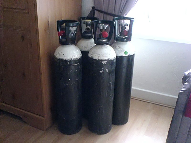
Week 51 – LOTT

“A Randomized Trial of Long-Term Oxygen for COPD with Moderate Desaturation”

by the Long-Term Oxygen Treatment Trial (LOTT) Research Group

N Engl J Med. 2016 Oct 27;375(17):1617-1627. [free full text]

The long-term treatment of severe resting hypoxemia (SpO2 < 89%) in COPD with supplemental oxygen has been a cornerstone of modern outpatient COPD management since its mortality benefit was demonstrated circa 1980. Subsequently, the utility of supplemental oxygen in COPD patients with moderate resting daytime hypoxemia (SpO2 89-93%) was investigated in trials in the 1990s; however, such trials were underpowered to assess mortality benefit. Ultimately, the LOTT trial was funded by the NIH and Centers for Medicare and Medicaid Services (CMS) primarily to determine if there was a mortality benefit to supplemental oxygen in COPD patients with moderate hypoxemia as well to analyze as numerous other secondary outcomes, such as hospitalization rates and exercise performance.

The LOTT trial was originally planned to enroll 3500 patients. However, after 7 months the trial had randomized only 34 patients, and mortality had been lower than anticipated. Thus in late 2009 the trial was redesigned to include broader inclusion criteria (now patients with exercise-induced hypoxemia could qualify) and the primary endpoint was broadened from mortality to a composite of time to first hospitalization or death.

The revised LOTT trial enrolled COPD patients with moderate resting hypoxemia (SpO2 89-93%) or moderate exercise-induced desaturation during the 6-minute walk test (SpO2 ≥ 80% for ≥ 5 minutes and < 90% for ≥ 10 seconds). Patients were randomized to either supplemental oxygen (24-hour oxygen if resting SpO2 89-93%, otherwise oxygen only during sleep and exercise if the desaturation occurred only during exercise) or to usual care without supplemental oxygen. Supplemental oxygen flow rate was 2 liters per minute and could be uptitrated by protocol among patients with exercise-induced hypoxemia. The primary outcome was time to composite of first hospitalization or death. Secondary outcomes included hospitalization rates, lung function, performance on 6-minute walk test, and quality of life.

368 patients were randomized to the supplemental-oxygen group and 370 to the no-supplemental-oxygen group. Of the supplemental-oxygen group, 220 patients were prescribed 24-hour oxygen support, and 148 were prescribed oxygen for use during exercise and sleep only. Median duration of follow-up was 18.4 months. Regarding the primary outcome, there was no group difference in time to death or first hospitalization (p = 0.52 by log-rank test). See Figure 1A. Furthermore, there were no treatment-group differences in the primary outcome among patients of the following pre-specified subgroups: type of oxygen prescription, “desaturation profile,” race, sex, smoking status, SpO2 nadir during 6-minute walk, FEV1, BODE  index, SF-36 physical-component score, BMI, or history of anemia. Patients with a COPD exacerbation in the 1-2 months prior to enrollment, age 71+ at enrollment, and those with lower Quality of Well-Being Scale score at enrollment all demonstrated benefit from supplemental O2, but none of these subgroup treatment effects were sustained when the analyses were adjusted for multiple comparisons. Regarding secondary outcomes, there were no treatment-group differences in rates of all-cause hospitalizations, COPD-related hospitalizations, or non-COPD-related hospitalizations, and there were no differences in change from baseline measures of quality of life, anxiety, depression, lung function, and distance achieved in 6-minute walk.

The LOTT trial presents compelling evidence that there is no significant benefit, mortality or otherwise, of oxygen supplementation in patients with COPD and either moderate hypoxemia at rest (SpO2 > 88%) or exercise-induced hypoxemia. Although this trial’s substantial redesign in its early course is noted, the trial still is our best evidence to date about the benefit (or lack thereof) of oxygen in this patient group. As acknowledged by the authors, the trial may have had significant selection bias in referral. (Many physicians did not refer specific patients for enrollment because “they were too ill or [were believed to have benefited] from oxygen.”) Another notable limitation of this study is that nocturnal oxygen saturation was not evaluated. The authors do note that “some patients with COPD and severe nocturnal desaturation might benefit from nocturnal oxygen supplementation.”

For further contemporary contextualization of the study, please see the excellent post at PulmCCM from 11/2016. Included in that post is a link to an overview and Q&A from the NIH regarding the LOTT study.

References / Additional Reading:
1. PulmCCM, “Long-term oxygen brought no benefits for moderate hypoxemia in COPD”
2. LOTT @ 2 Minute Medicine
3. LOTT @ ClinicalTrials.gov
4. McDonald, J.H. 2014. Handbook of Biological Statistics (3rd ed.). Sparky House Publishing, Baltimore, Maryland.
5. Centers for Medicare and Medicaid Services, “Certificate of Medical Necessity CMS-484– Oxygen”
6. Ann Am Thorac Soc. 2018 Dec;15(12):1369-1381. “Optimizing Home Oxygen Therapy. An Official American Thoracic Society Workshop Report.”

Summary by Duncan F. Moore, MD

Image Credit: Patrick McAleer, CC BY-SA 2.0 UK, via Wikimedia Commons
Week 32 – PneumA

“Comparison of 8 vs 15 Days of Antibiotic Therapy for Ventilator-Associated Pneumonia in Adults”

JAMA. 2003 November 19;290(19):2588-2598. [free full text]

Ventilator-associated pneumonia (VAP) is a frequent complication of mechanical ventilation and, prior to this study, few trials had addressed the optimal duration of antibiotic therapy in VAP. Thus, patients frequently received 14- to 21-day antibiotic courses. As antibiotic stewardship efforts increased and awareness grew of the association between prolonged antibiotic courses and the development of multidrug resistant (MDR) infections, more data were needed to clarify the optimal VAP treatment duration.

This 2003 trial by the PneumA Trial Group was the first large randomized trial to compare shorter (8-day) versus longer (15-day) treatment courses for VAP.

The noninferiority study, carried out in 51 French ICUs, enrolled intubated patients with clinical suspicion for VAP and randomized them to either 8 or 15 days of antimicrobials. Antimicrobial regimens were chosen by the treating clinician. 401 patients met eligibility criteria. 197 were randomized to the 8-day regimen. 204 patients were randomized to the 15-day regimen. Study participants were blinded to randomization assignment until day 8. Analysis was performed using an intention-to-treat model. The primary outcomes measured were death from any cause at 28 days, antibiotic-free days, and microbiologically documented pulmonary infection recurrence.

Study findings demonstrated a similar 28-day mortality in both groups (18.8% mortality in 8-day group vs. 17.2% in 15-day group, group difference 90% CI -3.7% to 6.9%). The 8-day group did not develop more recurrent infections (28.9% in 8-day group vs. 26.0% in 15-day group, group difference 90% CI -3.2% to 9.1%). The 8-day group did have more antibiotic-free days when measured at the 28-day point (13.1 in 8-day group vs. 8.7 in 15-day group, p<0.001). A subgroup analysis did show that more 8-day-group patients who had an initial infection with lactose-nonfermenting GNRs developed a recurrent pulmonary infection, so noninferiority was not established in this specific subgroup (40.6% recurrent GNR infection in 8-day group vs. 25.4% in 15-day group, group difference 90% CI 3.9% to 26.6%).

Implications/Discussion:
There is no benefit to prolonging VAP treatment to 15 days (except perhaps when Pseudomonas aeruginosa is suspected based on gram stain/culture data). Shorter courses of antibiotics for VAP treatment allow for less antibiotic exposure without increasing rates of recurrent infection or mortality.

The 2016 IDSA guidelines on VAP treatment recommend a 7-day course of antimicrobials for treatment of VAP (as opposed to a longer treatment course such as 8-15 days). These guidelines are based on the IDSA’s own large meta-analysis (of 10 randomized trials, including PneumA, as well as an observational study) which demonstrated that shorter courses of antibiotics (7 days) reduce antibiotic exposure and recurrent pneumonia due to MDR organisms without affecting clinical outcomes, such as mortality. Of note, this 7-day course recommendation also applies to treatment of lactose-nonfermenting GNRs, such as Pseudomonas.

When considering the PneumA trial within the context of the newest IDSA guidelines, we see that we now have over 15 years of evidence supporting the use of shorter VAP treatment courses.

Further Reading/References:
1. 2016 IDSA Guidelines for the Management of HAP/VAP
2. PneumA @ Wiki Journal Club
3. PulmCCM “IDSA Guidelines 2016: HAP, VAP & It’s the End of HCAP as We Know It (And I Feel Fine)”
4. PulmCrit “The siren’s call: Double-coverage for ventilator associated PNA”

Summary by Liz Novick, MD

Week 21 – PROSEVA

“Prone Positioning in Severe Acute Respiratory Distress Syndrome”
by the PROSEVA Study Group

N Engl J Med. 2013 June 6; 368(23):2159-2168 [free full text]

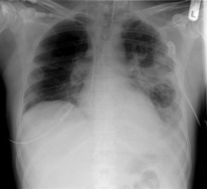
Prone positioning had been used for many years in ICU patients with ARDS in order to improve oxygenation. Per Dr. Sonti’s Georgetown Critical Care Top 40, the physiologic basis for benefit with proning lies in the idea that atelectatic regions of lung typically occur in the most dependent portion of an ARDS patient, with hyperinflation affecting the remaining lung. Periodic reversal of these regions via moving the patient from supine to prone and vice versa ensures no one region of the lung will have extended exposure to either atelectasis or overdistention. Although the oxygenation benefits have been long noted, the PROSEVA trial established mortality benefit.

Study patients were selected from 26 ICUs in France and 1 in Spain which had daily practice with prone positioning for at least 5 years. Inclusion criteria: ARDS patients intubated and ventilated <36hr with severe ARDS (defined as PaO2:FiO2 ratio < 150, PEEP > 5, and TV of about 6ml/kg of predicted body weight). (NB: by the Berlin definition for ARDS, severe ARDS is defined as PaO2:FiO2 ratio < 100.) Patients were either randomized to the intervention of proning within 36 hours of mechanical ventilation for at least 16 consecutive hours (n = 237) or to the control of being left in a semirecumbent (supine) position (n = 229). The primary outcome was mortality at day 28. Secondary outcomes included mortality at day 90, rate of successful extubation (no reintubation or use of noninvasive ventilation x48hr), time to successful extubation, length of stay in the ICU, complications, use of noninvasive ventilation, tracheotomy rate, number of days free from organ dysfunction, ventilator settings, measurements of ABG, and respiratory system mechanics during the first week after randomization.

At the time of randomization in the study, the majority of characteristics were similar between the two groups, although the authors noted differences in the SOFA score and the use of neuromuscular blockers and vasopressors. The supine group at baseline had a higher SOFA score indicating more severe organ failure, and also had higher rate of vasopressor usage. The prone group had a higher rate of usage of neuromuscular blockade. The primary outcome of 28 day mortality was significantly lower in the prone group than in the supine group, at 16.0% vs 32.8% (p < 0.001, NNT = 6.0). This mortality decrease was still statistically significant when adjusted for the SOFA score. Secondary outcomes were notable for a significantly higher rate of successful extubation in the prone group (hazard ratio 0.45; 95% CI 0.29-0.7, p < 0.001). Additionally, the PaO2:FiO2 ratio was significantly higher in the supine group, whereas the PEEP and FiO2 were significantly lower. The remainder of secondary outcomes were statistically similar.

PROSEVA showed a significant mortality benefit with early use of prone positioning in severe ARDS. This mortality benefit was considerably larger than that seen in past meta-analyses, which was likely due to this study selecting specifically for patients with severe disease as well as specifying longer prone-positioning sessions than employed in prior studies. Critics have noted the unexpected difference in baseline characteristics between the two arms of the study. While these critiques are reasonable, the authors mitigate at least some of these complaints by adjusting the mortality for the statistically significant differences. With such a radical mortality benefit it might be surprising that more patients are not proned at our institution. One reason is that relatively few of our patients have severe ARDS. Additionally, proning places a high demand on resources and requires a coordinated effort of multiple staff. All treatment centers in this study had specially-trained staff that had been performing proning on a daily basis for at least 5 years, and thus were very familiar with the process. With this in mind, we consider the use of proning in patients meeting criteria for severe ARDS.

References and further reading:
1. PROSEVA @ 2 Minute Medicine
2. PROSEVA @ Wiki Journal Club
3. PROSEVA @ Georgetown Critical Care Top 40, pages 8-9
4. Life in the Fastlane, Critical Care Compendium, “Prone Position and Mechanical Ventilation”
5. PulmCCM.org, “ICU Physiology in 1000 Words: The Hemodynamics of Prone”

Summary by Gordon Pelegrin, MD

Image Credit: by James Heilman, MD, CC BY-SA 3.0, via Wikimedia Commons

Week 17 – CURB-65

“Defining community acquired pneumonia severity on presentation to hospital: an international derivation and validation study”

Thorax. 2003 May;58(5):377-82. [free full text]

Community-acquired pneumonia (CAP) is frequently encountered by the admitting medicine team. Ideally, the patient’s severity at presentation and risk for further decompensation should determine the appropriate setting for further care, whether as an outpatient, on an inpatient ward, or in the ICU. At the time of this 2003 study, the predominant decision aid was the 20-variable Pneumonia Severity Index. The authors of this study sought to develop a simpler decision aid for determining the appropriate level of care at presentation.

The study examined the 30-day mortality rates of adults admitted for CAP via the ED at three non-US academic medical centers (data from three previous CAP cohort studies). 80% of the dataset was analyzed as a derivation cohort – meaning it was used to identify statistically significant, clinically relevant prognostic factors that allowed for mortality risk stratification. The resulting model was applied to the remaining 20% of the dataset (the validation cohort) in order to assess the accuracy of its predictive ability.

The following variables were integrated into the final model (CURB-65):

      1. Confusion
      2. Urea > 19mg/dL (7 mmol/L)
      3. Respiratory rate ≥ 30 breaths/min
      4. low Blood pressure (systolic BP < 90 mmHg or diastolic BP < 60 mmHg)
      5. age ≥ 65

1068 patients were analyzed. 821 (77%) were in the derivation cohort. 86% of patients received IV antibiotics, 5% were admitted to the ICU, and 4% were intubated. 30-day mortality was 9%. 9 of 11 clinical features examined in univariate analysis were statistically significant (see Table 2).

Ultimately, using the above-described CURB-65 model, in which 1 point is assigned for each clinical characteristic, patients with a CURB-65 score of 0 or 1 had 1.5% mortality, patients with a score of 2 had 9.2% mortality, and patients with a score of 3 or more had 22% mortality. Similar values were demonstrated in the validation cohort. Table 5 summarizes the sensitivity, specificity, PPVs, and NPVs of each CURB-65 score for 30-day mortality in both cohorts. As we would expect from a good predictive model, the sensitivity starts out very high and decreases with increasing score, while the specificity starts out very low and increases with increasing score. For the clinical application of their model, the authors selected the cut points of 1, 2, and 3 (see Figure 2).

In conclusion, CURB-65 is a simple 5-variable decision aid that is helpful in the initial stratification of mortality risk in patients with CAP.

The wide range of specificities and sensitivities at different values of the CURB-65 score makes it a robust tool for risk stratification. The authors felt that patients with a score of 0-1 were “likely suitable for home treatment,” patients with a score of 2 should have “hospital-supervised treatment,” and patients with score of  ≥ 3 had “severe pneumonia” and should be admitted (with consideration of ICU admission if score of 4 or 5).

Following the publication of the CURB-65 Score, the creator of the Pneumonia Severity Index (PSI) published a prospective cohort study of CAP that examined the discriminatory power (area under the receiver operating characteristic curve) of the PSI vs. CURB-65. His study found that the PSI “has a higher discriminatory power for short-term mortality, defines a greater proportion of patients at low risk, and is slightly more accurate in identifying patients at low risk” than the CURB-65 score.

Expert opinion at UpToDate prefers the PSI over the CURB-65 score based on its more robust base of confirmatory evidence. Of note, the author of the PSI is one of the authors of the relevant UpToDate article. In an important contrast from the CURB-65 authors, these experts suggest that patients with a CURB-65 score of 0 be managed as outpatients, while patients with a score of 1 and above “should generally be admitted.”

Further Reading/References:
1. Original publication of the PSI, NEJM (1997)
2. PSI vs. CURB-65 (2005)
3. CURB-65 @ Wiki Journal Club
4. CURB-65 @ 2 Minute Medicine
5. UpToDate, “CAP in adults: assessing severity and determining the appropriate level of care”

Summary by Duncan F. Moore, MD

Week 16 – National Lung Screening Trial (NLST)

“Reduced Lung-Cancer Mortality with Low-Dose Computed Tomographic Screening”

by the National Lung Cancer Screening Trial (NLST) Research Team

N Engl J Med. 2011 Aug 4;365(5):395-409 [free full text]

Despite a reduction in smoking rates in the United States, lung cancer remains the number one cause of cancer death in the United States as well as worldwide. Earlier studies of plain chest radiography for lung cancer screening demonstrated no benefit, and in 2002 the National Lung Screening Trial (NLST) was undertaken to determine whether then recent advances in CT technology could lead to an effective lung cancer screening method.

The study enrolled adults age 55-74 with 30+ pack-years of smoking (if former smokers, they must have quit within the past 15 years). Patients were randomized to either the intervention of three annual screenings for lung cancer with low-dose CT or to the comparator/control group to receive three annual screenings for lung cancer with PA chest radiograph. The primary outcome was mortality from lung cancer. Notable secondary outcomes were all-cause mortality and the incidence of lung cancer.

53,454 patients were randomized, and both groups had similar baseline characteristics. The low-dose CT group sustained 247 deaths from lung cancer per 100,000 person-years, whereas the radiography group sustained 309 deaths per 100,000 person-years. A relative reduction in rate of death by 20.0% was seen in the CT group (95% CI 6.8 – 26.7%, p = 0.004). The number needed to screen with CT to prevent one lung cancer death was 320. There were 1877 deaths from any cause in the CT group and 2000 deaths in the radiography group, so CT screening demonstrated a risk reduction of death from any cause of 6.7% (95% CI 1.2% – 13.6%, p = 0.02). Incidence of lung cancer in the CT group was 645 per 100,000 person-years and 941 per 100,000 person-years in the radiography group (RR 1.13, 95% CI 1.03 – 1.23).

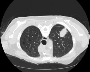
Lung cancer screening with low-dose CT scan in high-risk patients provides a significant mortality benefit. This trial was stopped early because the mortality benefit was so high. The benefit was driven by the reduction in deaths attributed to lung cancer, and when deaths from lung cancer were excluded from the overall mortality analysis, there was no significant difference among the two arms. Largely on the basis of this study, the 2013 USPSTF guidelines for lung cancer screening recommend annual low-dose CT scan in patients who meet NLST inclusion criteria. However, it must be noted that, even in the “ideal” circumstances of this trial performed at experienced centers, 96% of abnormal CT screening results in this trial were actually false positives. Of all positive results, 11% led to invasive studies.

Per UpToDate, since NSLT, there have been several European low-dose CT screening trials published. However, all but one (NELSON) appear to be underpowered to demonstrate a possible mortality reduction. Meta-analysis of all such RCTs could allow for further refinement in risk stratification, frequency of screening, and management of positive screening findings.

No randomized trial has ever demonstrated a mortality benefit of plain chest radiography for lung cancer screening. The Prostate, Lung, Colorectal, and Ovarian (PLCO) Cancer Screening Trial tested this modality vs. “community care,” and because the PLCO trial was ongoing at the time of creation of the NSLT, the NSLT authors trial decided to compare their intervention (CT) to plain chest radiography in case the results of plain chest radiography in PLCO were positive. Ultimately, they were not.

Further Reading:
1. USPSTF Guidelines for Lung Cancer Screening (2013)
2. NLST @ ClinicalTrials.gov
3. NLST @ Wiki Journal Club
4. NLST @ 2 Minute Medicine
5. UpToDate, “Screening for lung cancer”

Summary by Duncan F. Moore, MD

Image Credit: Yale Rosen, CC BY-SA 2.0, via Wikimedia Commons

Week 11 – Varenicline vs. Bupropion and Placebo for Smoking Cessation

“Varenicline, an α2β2 Nicotinic Acetylcholine Receptor Partial Agonist, vs Sustained-Release Bupropion and Placebo for Smoking Cessation”

JAMA. 2006 Jul 5;296(1):56-63. [free full text]

Assisting our patients in smoking cessation is a fundamental aspect of outpatient internal medicine. At the time of this trial, the only approved pharmacotherapies for smoking cessation were nicotine replacement therapy and bupropion. As the α2β2 nicotinic acetylcholine receptor (nAChR) was thought to be crucial to the reinforcing effects of nicotine, it was hypothesized that a partial agonist for this receptor could yield sufficient effect to satiate cravings and minimize withdrawal symptoms but also limit the reinforcing effects of exogenous nicotine. Thus Pfizer designed this large phase 3 trial to test the efficacy of its new α2β2 nAChR partial agonist varenicline (Chantix) against the only other non-nicotine pharmacotherapy at the time (bupropion) as well as placebo.

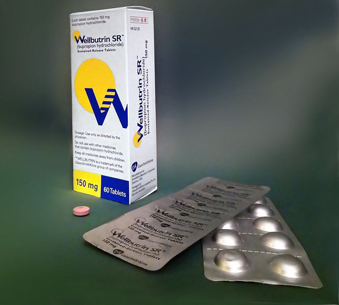
The trial enrolled adult smokers (10+ cigarettes per day) with fewer than three months of smoking abstinence in the past year (notable exclusion criteria included numerous psychiatric and substance use comorbidities). Patients were randomized to 12 weeks of treatment with either varenicline uptitrated by day 8 to 1mg BID, bupropion SR uptitrated by day 4 to 150mg BID, or placebo BID. Patients were also given a smoking cessation self-help booklet at the index visit and encouraged to set a quit date of day 8. Patients were followed at weekly clinic visits for the first 12 weeks (treatment duration) and then a mixture of clinic and phone visits for weeks 13-52. Non-smoking status during follow-up was determined by patient self-report combined with exhaled carbon monoxide < 10ppm. The primary endpoint was the 4-week continuous abstinence rate for study weeks 9-12 (as confirmed by exhaled CO level). Secondary endpoints included the continuous abstinence rate for weeks 9-24 and for weeks 9-52.

1025 patients were randomized. Compliance was similar among the three groups and the median duration of treatment was 84 days. Loss to follow-up was similar among the three groups. CO-confirmed continuous abstinence during weeks 9-12 was 44.0% among the varenicline group vs. 17.7% among the placebo group (OR 3.85, 95% CI 2.70–5.50, p < 0.001) vs. 29.5% among the bupropion group (OR vs. varenicline group 1.93, 95% CI 1.40–2.68, p < 0.001). (OR for bupropion vs. placebo was 2.00, 95% CI 1.38–2.89, p < 0.001.) Continuous abstinence for weeks 9-24 was 29.5% among the varenicline group vs. 10.5% among the placebo group (p < 0.001) vs. 20.7% among the bupropion group (p = 0.007). Continuous abstinence rates weeks 9-52 were 21.9% among the varenicline group vs. 8.4% among placebo group (p < 0.001) vs. 16.1% among the bupropion group (p = 0.057). Subgroup analysis of the primary outcome by sex did not yield significant differences in drug efficacy by sex.

This study demonstrated that varenicline was superior to both placebo and bupropion in facilitating smoking cessation at up to 24 weeks. At greater than 24 weeks, varenicline remained superior to placebo but was similarly efficacious as bupropion. This was a well-designed and executed large, double-blind, placebo- and active-treatment-controlled multicenter US trial. The trial was completed in April 2005 and a new drug application for varenicline (Chantix) was submitted to the FDA in November 2005. Of note, an “identically designed” (per this study’s authors), manufacturer-sponsored phase 3 trial was performed in parallel and reported very similar results in the in the same July 2006 issue of JAMA (PMID: 16820547) as the above study by Gonzales et al. These robust, positive-outcome pre-approval trials of varenicline helped the drug rapidly obtain approval in May 2006.

Per expert opinion at UpToDate, varenicline remains a preferred first-line pharmacotherapy for smoking cessation. Bupropion is a suitable, though generally less efficacious, alternative, particularly when the patient has comorbid depression. Per UpToDate, the recent (2016) EAGLES trial demonstrated that “in contrast to earlier concerns, varenicline and bupropion have no higher risk of associated adverse psychiatric effects than [nicotine replacement therapy] in smokers with comorbid psychiatric disorders.”

Further Reading/References:
1. This trial @ ClinicalTrials.gov
2. Sister trial: “Efficacy of varenicline, an alpha4beta2 nicotinic acetylcholine receptor partial agonist, vs placebo or sustained-release bupropion for smoking cessation: a randomized controlled trial.” JAMA. 2006 Jul 5;296(1):56-63.
3. Chantix FDA Approval Letter 5/10/2006
4. Rigotti NA. Pharmacotherapy for smoking cessation in adults. Post TW, ed. UpToDate. Waltham, MA: UpToDate Inc.
5. “Neuropsychiatric safety and efficacy of varenicline, bupropion, and nicotine patch in smokers with and without psychiatric disorders (EAGLES): a double-blind, randomised, placebo-controlled clinical trial.” Lancet. 2016 Jun 18;387(10037):2507-20.
6. 2 Minute Medicine: “Varenicline and bupropion more effective than varenicline alone for tobacco abstinence”
7. 2 Minute Medicine: “Varenicline safe for smoking cessation in patients with stable major depressive disorder”

Summary by Duncan F. Moore, MD

Image Credit: Сергей Фатеев, CC BY-SA 3.0, via Wikimedia Commons

Week 4 – ARDSNet

“Ventilation with Lower Tidal Volumes as Compared with Traditional Tidal Volumes for Acute Lung Injury and the Acute Respiratory Distress Syndrome”

by the Acute Respiratory Distress Syndrome Network (ARDSNet)

N Engl J Med. 2000 May 4;342(18):1301-8. [free full text]

Acute respiratory distress syndrome (ARDS) is an inflammatory and highly morbid lung injury found in many critically ill patients. In the 1990s, it was hypothesized that overdistention of aerated lung volumes and elevated airway pressures might contribute to the severity of ARDS, and indeed some work in animal models supported this theory. Prior to the ARDSNet study, four randomized trials had been conducted to investigate the possible protective effect of ventilation with lower tidal volumes, but their results were conflicting.

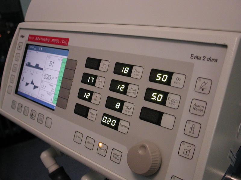
The ARDSNet study enrolled patients with ARDS (diagnosed within 36 hours) to either a lower initial tidal volume of 6ml/kg, downtitrated as necessary to maintain plateau pressure ≤ 30 cm H2O, or to the “traditional” therapy of an initial tidal volume of 12 ml/kg, downtitrated as necessary to maintain plateau pressure ≤ 50 cm of water. The primary outcomes were in-hospital mortality and ventilator-free days within the first 28 days. Secondary outcomes included number of days without organ failure, occurrence of barotrauma, and reduction in IL-6 concentration from day 0 to day 3.

861 patients were randomized before the trial was stopped early due to the increased mortality in the control arm noted during interim analysis. In-hospital mortality was 31.0% in the lower tidal volume group and 39.8% in the traditional tidal volume group (p = 0.007, NNT = 11.4). Ventilator free days were 12±11 in the lower tidal volume group vs. 10±11 in the traditional group (n = 0.007). The lower tidal volume group had more days without organ failure (15±11 vs. 12±11, p = 0.006). There was no difference in rates of barotrauma among the two groups. Decrease in IL-6 concentration between days 0 and 3 was greater in the low tidal volume group (p < 0.001), and IL-6 concentration at day 3 was lower in the low tidal volume group (p = 0.002).

In summary, low tidal volume ventilation decreases mortality in ARDS relative to “traditional” tidal volumes. The authors felt that this study confirmed the results of prior animal models and conclusively answered the question of whether or not low tidal volume ventilation provided a mortality benefit. In fact, in the years following, low tidal volume ventilation became the standard of care, and a robust body of literature followed this study to further delineate a “lung-protective strategy.” Critics of the study noted that, at the time of the study, the “traditional” (standard of care) tidal volume in ARDS was less than the 12 ml/kg used in the comparison arm. (Non-enrolled patients at the participating centers were receiving a mean tidal volume of 10.3 ml/kg.) Thus not only was the trial making a comparison to a faulty control, but it was also potentially harming patients in the control arm. An excellent summary of the ethical issues and debate regarding this specific issue and regarding control arms of RCTs in general can be found here.

Corresponding practice point from Dr. Sonti and Dr. Vinayak and their Georgetown Critical Care Top 40: “Low tidal volume ventilation is the standard of care in patients with ARDS (P/F < 300). Use ≤ 6 ml/kg predicted body weight, follow plateau pressures, and be cautious of mixed modes in which you set a tidal volume but the ventilator can adjust and choose a larger one.”

PulmCCM is an excellent blog, and they have a nice page reviewing this topic and summarizing some of the research and guidelines that have followed.

Further Reading/References:
1. ARDSNet @ Wiki Journal Club
2. ARDSNet @ 2 Minute Medicine
3. PulmCCM “Mechanical Ventilation in ARDS: Research Update”
4. Georgetown Critical Care Top 40, page 6
5. PulmCCM “In ARDS, substandard ventilator care is the norm, not the exception.” 2017.

Summary by Duncan F. Moore, MD

Week 40 – PROSEVA

Prone Positioning in Severe Acute Respiratory Distress Syndrome
by the PROSEVA Study Group

N Engl J Med. 2013 June 6; 368(23):2159-2168 [free full text]

Prone positioning had been used for many years in ICU patients with ARDS in order to improve oxygenation. Per Dr. Sonti’s Georgetown Critical Care Top 40, the physiologic basis for benefit with proning lies in the idea that atelectatic regions of lung typically occur in the most dependent portion of an ARDS patient, with hyperinflation affecting the remaining lung. Periodic reversal of these regions via moving the patient from supine to prone and vice versa ensures no one region of the lung will have extended exposure to either atelectasis or overdistention. Although the oxygenation benefits have been long noted, the PROSEVA trial established mortality benefit.

Study patients were selected from 26 ICUs in France and 1 in Spain which had daily practice with prone positioning for at least 5 years. Inclusion criteria: ARDS patients intubated and ventilated <36hr with severe ARDS (defined as PaO2:FiO2 ratio <150, PEEP>5, and TV of about 6ml/kg of predicted body weight). (NB: by the Berlin definition for ARDS, severe ARDS is defined as PaO2:FiO2 ratio <100.) Patients were either randomized to the intervention of proning within 36 hours of mechanical ventilation for at least 16 consecutive hours (N=237) or to the control of being left in a semirecumbent (supine) position (N=229). The primary outcome was mortality at day 28. Secondary outcomes included mortality at day 90, rate of successful extubation (no reintubation or use of noninvasive ventilation x48hr), time to successful extubation, length of stay in the ICU, complications, use of noninvasive ventilation, tracheotomy rate, number of days free from organ dysfunction, ventilator settings, measurements of ABG, and respiratory system mechanics during the first week after randomization.

At the time of randomization in the study, the majority of characteristics were similar between the two groups, although the authors noted differences in the SOFA score and the use of neuromuscular blockers and vasopressors. The supine group at baseline had a higher SOFA score indicating more severe organ failure, and also had higher rate of vasopressor usage. The prone group had a higher rate of usage of neuromuscular blockade. The primary outcome of 28 day mortality was significantly lower in the prone group than in the supine group, at 16.0% vs 32.8% (p < 0.001, NNT = 6.0). This mortality decrease was still statistically significant when adjusted for the SOFA score. Secondary outcomes were notable for a significantly higher rate of successful extubation in the prone group (hazard ratio 0.45; 95% CI 0.29-0.7, p < 0.001). Additionally, the PaO2:FiO2 ratio was significantly higher in the supine group, whereas the PEEP and FiO2 were significantly lower. The remainder of secondary outcomes were statistically similar.

PROSEVA showed a significant mortality benefit with early use of prone positioning in severe ARDS. This mortality benefit was considerably larger than that seen in past meta-analyses, which was likely due to this study selecting specifically for patients with severe disease as well as specifying longer prone-positioning sessions than employed in prior studies. Critics have noted the unexpected difference in baseline characteristics between the two arms of the study. While these critiques are reasonable, the authors mitigate at least some of these complaints by adjusting the mortality for the statistically significant differences. With such a radical mortality benefit it might be surprising that more patients are not proned at our institution. One reason is that relatively few of our patients have severe ARDS. Additionally, proning places a high demand on resources and requires a coordinated effort of multiple staff. All treatment centers in this study had specially-trained staff that had been performing proning on a daily basis for at least 5 years, and thus were very familiar with the process. With this in mind, we consider the use of proning in patients meeting criteria for severe ARDS.

References and further reading:
1. PROSEVA @ 2 Minute Medicine
2. PROSEVA @ Wiki Journal Club
3. PROSEVA @ Georgetown Critical Care Top 40, pages 8-9
4. Life in the Fastlane, Critical Care Compendium, “Prone Position and Mechanical Ventilation”
5. PulmCCM.org, “ICU Physiology in 1000 Words: The Hemodynamics of Prone”

Summary by Gordon Pelegrin, MD

Image Credit: by James Heilman, MD, CC BY-SA 3.0, via Wikimedia Commons

Week 37 – LOTT

“A Randomized Trial of Long-Term Oxygen for COPD with Moderate Desaturation”

by the Long-Term Oxygen Treatment Trial (LOTT) Research Group

N Engl J Med. 2016 Oct 27;375(17):1617-1627. [free full text]

The long-term treatment of severe resting hypoxemia (SpO2 < 89%) in COPD with supplemental oxygen has been a cornerstone of modern outpatient COPD management since its mortality benefit was demonstrated circa 1980. Subsequently, the utility of supplemental oxygen in COPD patients with moderate resting daytime hypoxemia (SpO2 89-93%) was investigated in trials in the 1990s; however, such trials were underpowered to assess mortality benefit. Ultimately, the LOTT trial was funded by the NIH and Centers for Medicare and Medicaid Services (CMS) primarily to determine if there was a mortality benefit to supplemental oxygen in COPD patients with moderate hypoxemia as well to analyze as numerous other secondary outcomes, such as hospitalization rates and exercise performance.

The LOTT trial was originally planned to enroll 3500 patients. However, after 7 months the trial had randomized only 34 patients, and mortality had been lower than anticipated. Thus in late 2009 the trial was redesigned to include broader inclusion criteria (now patients with exercise-induced hypoxemia could qualify) and the primary endpoint was broadened from mortality to a composite of time to first hospitalization or death.

The revised LOTT trial enrolled COPD patients with moderate resting hypoxemia (SpO2 89-93%) or moderate exercise-induced desaturation during the 6-minute walk test (SpO2 ≥ 80% for ≥ 5 minutes and < 90% for ≥ 10 seconds). Patients were randomized to either supplemental oxygen (24-hour oxygen if resting SpO2 89-93%, otherwise oxygen only during sleep and exercise if the desaturation occurred only during exercise) or to usual care without supplemental oxygen. Supplemental oxygen flow rate was 2 liters per minute and could be uptitrated by protocol among patients with exercise-induced hypoxemia. The primary outcome was time to composite of first hospitalization or death. Secondary outcomes included hospitalization rates, lung function, performance on 6-minute walk test, and quality of life.

368 patients were randomized to the supplemental-oxygen group and 370 to the no-supplemental-oxygen group. Of the supplemental-oxygen group, 220 patients were prescribed 24-hour oxygen support, and 148 were prescribed oxygen for use during exercise and sleep only. Median duration of follow-up was 18.4 months. Regarding the primary outcome, there was no group difference in time to death or first hospitalization (p = 0.52 by log-rank test). See Figure 1A. Furthermore, there were no treatment-group differences in the primary outcome among patients of the following pre-specified subgroups: type of oxygen prescription, “desaturation profile,” race, sex, smoking status, SpO2 nadir during 6-minute walk, FEV1, BODE  index, SF-36 physical-component score, BMI, or history of anemia. Patients with a COPD exacerbation in the 1-2 months prior to enrollment, age 71+ at enrollment, and those with lower Quality of Well-Being Scale score at enrollment all demonstrated benefit from supplemental O2, but none of these subgroup treatment effects were sustained when the analyses were adjusted for multiple comparisons. Regarding secondary outcomes, there were no treatment-group differences in rates of all-cause hospitalizations, COPD-related hospitalizations, or non-COPD-related hospitalizations, and there were no differences in change from baseline measures of quality of life, anxiety, depression, lung function, and distance achieved in 6-minute walk.

The LOTT trial presents compelling evidence that there is no significant benefit, mortality or otherwise, of oxygen supplementation in patients with COPD and either moderate hypoxemia at rest (SpO2 > 88%) or exercise-induced hypoxemia. Although this trial’s substantial redesign in its early course is noted, the trial still is our best evidence to date about the benefit (or lack thereof) of oxygen in this patient group. As acknowledged by the authors, the trial may have had significant selection bias in referral. (Many physicians did not refer specific patients for enrollment because “they were too ill or [were believed to have benefited] from oxygen.”) Another notable limitation of this study is that nocturnal oxygen saturation was not evaluated. The authors do note that “some patients with COPD and severe nocturnal desaturation might benefit from nocturnal oxygen supplementation.”

For further contemporary contextualization of the study, please see the excellent post at PulmCCM from 11/2016. Included in that post is a link to an overview and Q&A from the NIH regarding the LOTT study.

References / Additional Reading:
1. PulmCCM, “Long-term oxygen brought no benefits for moderate hypoxemia in COPD”
2. LOTT @ 2 Minute Medicine
3. LOTT @ ClinicalTrials.gov
4. McDonald, J.H. 2014. Handbook of Biological Statistics (3rd ed.). Sparky House Publishing, Baltimore, Maryland.
5. Centers for Medicare and Medicaid Services, “Certificate of Medical Necessity CMS-484– Oxygen”
6. Ann Am Thorac Soc. 2018 Dec;15(12):1369-1381. “Optimizing Home Oxygen Therapy. An Official American Thoracic Society Workshop Report.”

Summary by Duncan F. Moore, MD

Image Credit: Patrick McAleer, CC BY-SA 2.0 UK, via Wikimedia Commons
Week 33 – Varenicline vs. Bupropion and Placebo for Smoking Cessation

“Varenicline, an α2β2 Nicotinic Acetylcholine Receptor Partial Agonist, vs Sustained-Release Bupropion and Placebo for Smoking Cessation”

JAMA. 2006 Jul 5;296(1):56-63. [free full text]

Assisting our patients in smoking cessation is a fundamental aspect of outpatient internal medicine. At the time of this trial, the only approved pharmacotherapies for smoking cessation were nicotine replacement therapy and bupropion. As the α2β2 nicotinic acetylcholine receptor (nAChR) was thought to be crucial to the reinforcing effects of nicotine, it was hypothesized that a partial agonist for this receptor could yield sufficient effect to satiate cravings and minimize withdrawal symptoms but also limit the reinforcing effects of exogenous nicotine. Thus Pfizer designed this large phase 3 trial to test the efficacy of its new α2β2 nAChR partial agonist varenicline (Chantix) against the only other non-nicotine pharmacotherapy at the time (bupropion) as well as placebo.

The trial enrolled adult smokers (10+ cigarettes per day) with fewer than three months of smoking abstinence in the past year (notable exclusion criteria included numerous psychiatric and substance use comorbidities). Patients were randomized to 12 weeks of treatment with either varenicline uptitrated by day 8 to 1mg BID, bupropion SR uptitrated by day 4 to 150mg BID, or placebo BID. Patients were also given a smoking cessation self-help booklet at the index visit and encouraged to set a quit date of day 8. Patients were followed at weekly clinic visits for the first 12 weeks (treatment duration) and then a mixture of clinic and phone visits for weeks 13-52. Non-smoking status during follow-up was determined by patient self-report combined with exhaled carbon monoxide < 10ppm. The primary endpoint was the 4-week continuous abstinence rate for study weeks 9-12 (as confirmed by exhaled CO level). Secondary endpoints included the continuous abstinence rate for weeks 9-24 and for weeks 9-52.

1025 patients were randomized. Compliance was similar among the three groups and the median duration of treatment was 84 days. Loss to follow-up was similar among the three groups. CO-confirmed continuous abstinence during weeks 9-12 was 44.0% among the varenicline group vs. 17.7% among the placebo group (OR 3.85, 95% CI 2.70–5.50, p < 0.001) vs. 29.5% among the bupropion group (OR vs. varenicline group 1.93, 95% CI 1.40–2.68, p < 0.001). (OR for bupropion vs. placebo was 2.00, 95% CI 1.38–2.89, p < 0.001.)  Continuous abstinence for weeks 9-24 was 29.5% among the varenicline group vs. 10.5% among the placebo group (p < 0.001) vs. 20.7% among the bupropion group (p = 0.007). Continuous abstinence rates weeks 9-52 were 21.9% among the varenicline group vs. 8.4% among placebo group (p < 0.001) vs. 16.1% among the bupropion group (p = 0.057). Subgroup analysis of the primary outcome by sex did not yield significant differences in drug efficacy by sex.

This study demonstrated that varenicline was superior to both placebo and bupropion in facilitating smoking cessation at up to 24 weeks. At greater than 24 weeks, varenicline remained superior to placebo but was similarly efficacious as bupropion. This was a well-designed and executed large, double-blind, placebo- and active-treatment-controlled multicenter US trial. The trial was completed in April 2005 and a new drug application for varenicline (Chantix) was submitted to the FDA in November 2005. Of note, an “identically designed” (per this study’s authors), manufacturer-sponsored phase 3 trial was performed in parallel and reported very similar results in the in the same July 2006 issue of JAMA (PMID: 16820547) as the above study by Gonzales et al. These robust, positive-outcome pre-approval trials of varenicline helped the drug rapidly obtain approval in May 2006.

Per expert opinion at UpToDate, varenicline remains a preferred first-line pharmacotherapy for smoking cessation. Bupropion is a suitable, though generally less efficacious, alternative, particularly when the patient has comorbid depression. Per UpToDate, the recent (2016) EAGLES trial demonstrated that “in contrast to earlier concerns, varenicline and bupropion have no higher risk of associated adverse psychiatric effects than [nicotine replacement therapy] in smokers with comorbid psychiatric disorders.”

Further Reading/References:
1. This trial @ ClinicalTrials.gov
2. Sister trial: “Efficacy of varenicline, an alpha4beta2 nicotinic acetylcholine receptor partial agonist, vs placebo or sustained-release bupropion for smoking cessation: a randomized controlled trial.” JAMA. 2006 Jul 5;296(1):56-63.
3. Chantix FDA Approval Letter 5/10/2006
4. Rigotti NA. Pharmacotherapy for smoking cessation in adults. Post TW, ed. UpToDate. Waltham, MA: UpToDate Inc. [https://www.uptodate.com/contents/pharmacotherapy-for-smoking-cessation-in-adults] (Accessed on February 16, 2019).
5. “Neuropsychiatric safety and efficacy of varenicline, bupropion, and nicotine patch in smokers with and without psychiatric disorders (EAGLES): a double-blind, randomised, placebo-controlled clinical trial.” Lancet. 2016 Jun 18;387(10037):2507-20.
6. 2 Minute Medicine: “Varenicline and bupropion more effective than varenicline alone for tobacco abstinence”
7. 2 Minute Medicine: “Varenicline safe for smoking cessation in patients with stable major depressive disorder”

Summary by Duncan F. Moore, MD

Image Credit: Сергей Фатеев, CC BY-SA 3.0, via Wikimedia Commons