Week 44 – National Lung Screening Trial (NLST)

“Reduced Lung-Cancer Mortality with Low-Dose Computed Tomographic Screening”

by the National Lung Cancer Screening Trial (NLST) Research Team

N Engl J Med. 2011 Aug 4;365(5):395-409 [free full text]

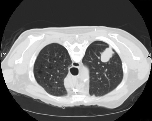
Despite a reduction in smoking rates in the United States, lung cancer remains the number one cause of cancer death in the United States as well as worldwide. Earlier studies of plain chest radiography for lung cancer screening demonstrated no benefit, and in 2002 the National Lung Screening Trial (NLST) was undertaken to determine whether then recent advances in CT technology could lead to an effective lung cancer screening method.

The study enrolled adults age 55-74 with 30+ pack-years of smoking (if former smokers, they must have quit within the past 15 years). Patients were randomized to either the intervention of three annual screenings for lung cancer with low-dose CT or to the comparator/control group to receive three annual screenings for lung cancer with PA chest radiograph. The primary outcome was mortality from lung cancer. Notable secondary outcomes were all-cause mortality and the incidence of lung cancer.

53,454 patients were randomized, and both groups had similar baseline characteristics. The low-dose CT group sustained 247 deaths from lung cancer per 100,000 person-years, whereas the radiography group sustained 309 deaths per 100,000 person-years. A relative reduction in rate of death by 20.0% was seen in the CT group (95% CI 6.8 – 26.7%, p = 0.004). The number needed to screen with CT to prevent one lung cancer death was 320. There were 1877 deaths from any cause in the CT group and 2000 deaths in the radiography group, so CT screening demonstrated a risk reduction of death from any cause of 6.7% (95% CI 1.2% – 13.6%, p = 0.02). Incidence of lung cancer in the CT group was 645 per 100,000 person-years and 941 per 100,000 person-years in the radiography group (RR 1.13, 95% CI 1.03 – 1.23).

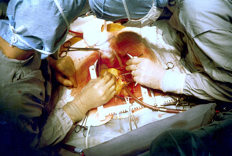
Lung cancer screening with low-dose CT scan in high-risk patients provides a significant mortality benefit. This trial was stopped early because the mortality benefit was so high. The benefit was driven by the reduction in deaths attributed to lung cancer, and when deaths from lung cancer were excluded from the overall mortality analysis, there was no significant difference among the two arms. Largely on the basis of this study, the 2013 USPSTF guidelines for lung cancer screening recommend annual low-dose CT scan in patients who meet NLST inclusion criteria. However, it must be noted that, even in the “ideal” circumstances of this trial performed at experienced centers, 96% of abnormal CT screening results in this trial were actually false positives. Of all positive results, 11% led to invasive studies.

Per UpToDate, since NSLT, there have been several European low-dose CT screening trials published. However, all but one (NELSON) appear to be underpowered to demonstrate a possible mortality reduction. Meta-analysis of all such RCTs could allow for further refinement in risk stratification, frequency of screening, and management of positive screening findings.

No randomized trial has ever demonstrated a mortality benefit of plain chest radiography for lung cancer screening. The Prostate, Lung, Colorectal, and Ovarian (PLCO) Cancer Screening Trial tested this modality vs. “community care,” and because the PLCO trial was ongoing at the time of creation of the NSLT, the NSLT authors trial decided to compare their intervention (CT) to plain chest radiography in case the results of plain chest radiography in PLCO were positive. Ultimately, they were not.

Further Reading:
1. USPSTF Guidelines for Lung Cancer Screening (2013)
2. NLST @ ClinicalTrials.gov
3. NLST @ Wiki Journal Club
4. NLST @ 2 Minute Medicine
5. UpToDate, “Screening for lung cancer”

Summary by Duncan F. Moore, MD

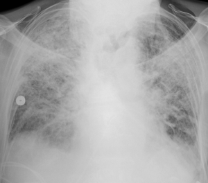
Image Credit: Yale Rosen, CC BY-SA 2.0, via Wikimedia Commons

Week 43 – FREEDOM

“Strategies for Multivessel Revascularization in Patients with Diabetes”

by the FREEDOM (Future Revascularization Evaluation in Patients with Diabetes Mellitus: Optimal Management of Multivessel Disease) Trial investigators

N Engl J Med. 2012 Dec 20;367(25):2375-84. [free full text]

Previous studies, such as the 1996 BARI trial), have demonstrated that patients who have multivessel coronary artery disease (CAD) and diabetes mellitus (DM) and who received coronary artery bypass grafting (CABG) surgery lived longer than patients undergoing balloon angioplasty. However, since that publication, percutaneous coronary intervention (PCI) technology advanced significantly. Prior to the publication of FREEDOM in 2012, there had only been small, underpowered studies comparing PCI with drug-eluting stent (DES) to CABG. FREEDOM was powered appropriately to discover superiority of revascularization strategy (PCI with DES vs. CABG) in patients with DM and multivessel CAD.

Population:

Inclusion criteria:

      • 18 years or older
      • Diabetes mellitus – defined by American Diabetes Association
      • Multivessel Coronary Artery Disease
        • > 70% stenosis (angiographically confirmed)
        • 2 or more epicardial vessels
        • 2 or more coronary-artery territories

Selected exclusion criteria:

      • NYHA Class III-IV heart failure
      • Prior CABG, valve surgery, or PCI (< 6 months)
      • Prior significant bleed (< 6 months)
      • Left main stenosis ≥ 50%


Design:
Patients meeting criteria were assigned 1:1 into PCI with first-generation paclitaxel-eluting stent (51%) or sirolimus-eluting stent (43%) versus CABG. The PCI group was placed on aspirin and clopidogrel for dual antiplatelet therapy (DAPT) for at least 12 months. For the CABG group, arterial revascularization was encouraged. The mean SYNTAX score (tool used to score complexity of CAD) was 26.2 and did not significantly differ between groups. Guideline-driven targets for lowering medical risk factors were used: LDL <70, BP <130/80, HgbA1c <7. Minimum follow-up was 2 years.


Outcomes:

Primary: Composite of death from any cause, non-fatal myocardial infarction (MI), and non-fatal stroke

Secondary

      1. Rate of major adverse cardiovascular and cerebrovascular events at 30 days and 12 months
      2. Repeat revascularization
      3. Annual all-cause mortality
      4. Annual cardiovascular mortality


Results:
953 patients and 947 patients were randomized into the PCI and CABG groups, respectively. At 5 years, the primary outcome (combined death, MI, or stroke) occurred in 200 of the PCI group and 146 of the CABG group (26.6% vs 18.7%, p = 0.005). The curves started diverging at 2 years. All-cause mortality was higher in the PCI group versus the CABG group (16.3% vs 10.9%, p = 0.049). Regarding secondary outcomes, 13.9% of patients in the PCI group had a repeat MI versus 6.0% in the CABG group (p < 0.001). There were fewer strokes in the PCI group than in the CABG group (2.4% vs 5.2%, p = 0.03). There was no statistically significant difference between study groups regarding cardiovascular death (10.9% vs 6.8%, p = 0.12).

At 5 years, the analysis of outcomes according to category of SYNTAX score (≤ 22, 23 to 32, ≥ 33) showed no significant subgroup interaction (p = 0.58).

Regarding safety, major bleeding between the two groups at 30 days was 0.02% for PCI vs 0.04% for CABG (p = 0.13). The incidence of acute renal failure requiring hemodialysis was observed in one patient in the PCI group and eight patients in the CABG group (p = 0.02)

Implication/Discussion:
The BARI Trial (1996) was the first trial to show that patients with DM and multivessel CAD derive mortality benefit from bypass grafting over PCI with balloon angioplasty. Furthermore, the BARI 2D (2009) trial demonstrated this benefit of bypass grafting over PCI with bare metal stents (BMS). At the time of the FREEDOM Trial, there had not been a randomized comparison of CABG versus PCI with newer technology and first-generation paclitaxel/sirolimus DES. In this study, CABG showed a 5.3% absolute reduction in all-cause mortality over PCI as well decreased rates of MI and repeat revascularization. CABG was associated with a mild absolute increase in stroke (2.8%). However, this mild increased stroke risk is consistent with most other comparative trials of the two treatment strategies. There was no statistical difference in major bleeding between the two groups.

CABG is likely better than PCI for various reasons. For one, diabetic arteries are affected diffusely and tend to have more extensive atherosclerotic disease compared to those without diabetes, so the likelihood of successful PCI alone is low. Many suspected that with advancement in PCI (i.e. DES) that the BARI data would become irrelevant. However, CABG continued to show benefit despite the technological advancements of drug-eluting stents and PCI. Improvement in surgical technique as well as the use of arterial revascularization (i.e. internal mammary artery) helped maintain superior outcomes with CABG compared to PCI.

The study was limited by the fact that due to low numbers, the subgroup analysis (i.e. SYNTAX scores) was not appropriately powered for statistical significance. Further, the study was not blinded, and patients may have been treated differently on the basis of their surgical procedure. Also, there was variability of STYNAX scores between the study groups, but this circumstance was thought to reflect real world heterogeneity.

Bottom Line:
CABG was superior to PCI with DES in patients with DM and multivessel CAD in that it significantly reduced rates of death and MI despite a small increased risk of stroke.

Further Reading/References:
1. BARI Trial @ NEJM
2. BARI 2D Trial @ NEJM
3. ACCF/AHA 2011 Guideline for Coronary Artery Bypass Graft Surgery
4. FREEDOM @ Wiki Journal Club
5. FREEDOM @ 2 Minute Medicine
5. FREEDOM @ Visualmed

Summary by Patrick Miller, MD.

Image Credit: Jerry Hecht, US Public Domain, via Wikimedia Commons

Week 42 – BeSt

“Clinical and Radiographic Outcomes of Four Different Treatment Strategies in Patients with Early Rheumatoid Arthritis (the BeSt Study).”

Arthritis & Rheumatism. 2005 Nov;52(11):3381-3390. [free full text]

Rheumatoid arthritis (RA) is among the most prevalent of the rheumatic diseases with a lifetime prevalence of 3.6% in women and 1.7% in men [1]. It is a chronic, systemic, inflammatory autoimmune disease of variable clinical course that can severely impact physical functional status and even mortality. Over the past 30 years, as the armamentarium of therapies for RA has exploded, there has been increased debate about the ideal initial therapy. The BeSt (Dutch: Behandel-Strategieën “treatment strategies”) trial was designed to compare, according to the authors, four of “the most frequently used and discussed strategies.” Regimens incorporating traditional disease-modifying antirheumatic drugs (DMARDs), such as methotrexate, and newer therapies, such as TNF-alpha inhibitors, were compared directly.

The trial enrolled 508 DMARD-naïve patients with early rheumatoid arthritis. Pertinent exclusion criteria included history of cancer and pre-existing laboratory abnormalities or comorbidities (e.g. elevated creatinine or ALT, alcohol abuse, pregnancy or desire to conceive, etc.) that would preclude the use of various DMARDs. Patients were randomized to one of four treatment groups. Within each regimen, the Disease Activity Score in 44 joints (DAS-44) was assessed q3 months, and, if > 2.4, the medication regimen was uptitrated to the next step within the treatment group.

Four Treatment Groups

  1. Sequential monotherapy: methotrexate (MTX) 15mg/week, uptitrated PRN to 25-30mg/week. If insufficient control, the following sequence was pursued: sulfasalazine (SSZ) monotherapy, leflunomide monotherapy, MTX + infliximab, gold with methylprednisolone, MTX + cyclosporin A (CSA) + prednisone
  2. Step-up combination therapy: MTX 15mg/week, uptitrated PRN to 25-30mg/week. If insufficient control, SSZ was added, followed by hydroxychloroquine (HCQ), followed by prednisone. If patients failed to respond to those four drugs, they were switched to MTX + infliximab, then MTX + CSA + prednisone, and finally to leflunomide.
  3. Initial combination therapy with tapered high-dose prednisone: MTX 7.5 mg/week + SSZ 2000 mg/day + prednisone 60mg/day (tapered in 7 weeks to 7.5 mg/day). If insufficient control, MTX was uptitrated to 25-30 mg/week. Next, combination would be switched to MTX + CSA + prednisone, then MTX + infliximab, then leflunomide monotherapy, gold with methylprednisolone, and finally azathioprine with prednisone.
  4. Initial combination therapy with infliximab: MTX 25-30 mg/week + infliximab 3 mg/kg at weeks 0, 2, 6, and q8 weeks thereafter. There was a protocol for infliximab-dose uptitration starting at 3 months. If insufficient control on MTX and infliximab 10 mg/kg, patients were switched to SSZ, then leflunomide, then MTX + CSA + prednisone, then gold + methylprednisolone, and finally AZA with prednisone.

Once clinical response was adequate for at least 6 months, there was a protocol for tapering the drug regimen.

The primary endpoints were: 1) functional ability per the Dutch version of the Health Assessment Questionnaire (D-HAQ), collected by a blinded research nurse q3 months and 2) radiographic joint damage per the modified Sharp/Van der Heijde score (SHS). Pertinent secondary outcomes included DAS-44 score and laboratory evidence of treatment toxicity.

At randomization, enrolled RA patients had a median duration of symptoms of 23 weeks and median duration since diagnosis of RA of 2 weeks. Mean DAS-44 was 4.4 ± 0.9. 72% of patients had erosive disease. Mean D-HAQ score at 3 months was 1.0 in groups 1 and 2 and 0.6 in groups 3 and 4 (p < 0.001 for groups 1 and 2 vs. groups 3 and 4; paired tests otherwise insignificant). Mean D-HAQ at 1 year was 0.7 in groups 1 and 2 and 0.5 in groups 3 and 4 (p = 0.010 for group 1 vs. group 3, p = 0.003 for group 1 vs. group 4; paired tests otherwise insignificant). At 1 year, patients in group 3 or 4 had less radiographic progression in joint damage per SHS than patients in group 1 or 2. Median increases in SHS were 2.0, 2.5., 1.0, and 0.5 in groups 1-4, respectively (p = 0.003 for group 1 vs. group 3, p < 0.001 for group 1 versus group 4, p = 0.007 for group 2 vs. group 3, p < 0.001 for group 2 vs. group 4). Regarding DAS-44 score: low disease activity (DAS-44 ≤ 2.4) at 1 year was reached in 53%, 64%, 71%, 74% of groups 1-4, respectively (p = 0.004 for group 1 vs. group 3, p = 0.001 for group 1 vs. group 4, p not significant for other comparisons). There were no group differences in prevalence of adverse effects.

Overall, among patients with early RA, initial combination therapy that included either prednisone (group 3) or infliximab (group 4) resulted in better functional and radiographic improvement than did initial therapy with sequential monotherapy (group 1) or step-up combination therapy (group 2). In the discussion, the authors note that given the treatment group differences in radiographic progression of disease, “starting therapy with a single DMARD would be a missed opportunity in a considerable number of patients.” Contemporary commentary by Weisman notes that “the authors describe both an argument and a counterargument arising from their observations: aggressive treatment with combinations of expensive drugs would ‘overtreat’ a large proportion of patients, yet early suppression of disease activity may have an important influence on subsequent long‐term disability and damage.”

Fourteen years later, it is a bit difficult to place the specific results of this trial in our current practice. Its trial design is absolutely byzantine and compares the 1-year experience of a variety of complex protocols that theoretically have substantial eventual potential overlap. Furthermore, it is difficult to assess if the relatively small group differences in symptom (D-HAQ) and radiographic (SHS) scales were truly clinically significant even if they were statistically significant. The American College of Rheumatology 2015 Guideline for the Treatment of Rheumatoid Arthritis synthesized the immense body of literature that came before and after the BeSt study and ultimately gave a variety of conditional statements about the “best practice” treatment of symptomatic early RA. (See Table 2 on page 8.) The recommendations emphasized DMARD monotherapy as the initial strategy but in the specific setting of a treat-to-target strategy. They also recommended escalation to combination DMARDs or biologics in patients with moderate or high disease activity despite DMARD monotherapy.

References / Additional Reading:
1. “The lifetime risk of adult-onset rheumatoid arthritis and other inflammatory autoimmune rheumatic diseases.” Arthritis Rheum. 2011 Mar;63(3):633-9. [https://www.ncbi.nlm.nih.gov/pubmed/21360492]
2. BeSt @ Wiki Journal Club
3. “Progress toward the cure of rheumatoid arthritis? The BeSt study.” Arthritis Rheum. 2005 Nov;52(11):3326-32.
4. “Review: treat to target in rheumatoid arthritis: fact, fiction, or hypothesis?” Arthritis Rheumatol. 2014 Apr;66(4):775-82. [https://www.ncbi.nlm.nih.gov/pubmed/24757129]
5. “2015 American College of Rheumatology Guideline for the Treatment of Rheumatoid Arthritis” Arthritis Rheumatol. 2016 Jan;68(1):1-26
6. RheumDAS calculator

Summary by Duncan F. Moore, MD

Image Credit: Braegel, CC BY 3.0, via Wikimedia Commons

Week 41 – Transfusion Strategies for Upper GI Bleeding

“Transfusion Strategies for Acute Upper Gastrointestinal Bleeding”

N Engl J Med. 2013 Jan 3;368(1):11-21. [free full text]

A restrictive transfusion strategy of 7 gm/dL was established following the previously discussed 1999 TRICC trial. Notably, both TRICC and its derivative study TRISS excluded patients who had an active bleed. In 2013, Villanueva et al. performed a study to establish whether there was benefit to a restrictive transfusion strategy in patients with acute upper GI bleeding.

The study enrolled consecutive adults presenting to a single center in Spain with hematemesis (or bloody nasogastric aspirate), melena, or both. Notable exclusion criteria included: a clinical Rockall score* of 0 with a hemoglobin level higher than 12g/dL, massive exsanguinating bleeding, lower GIB, patient refusal of blood transfusion, ACS, stroke/TIA, transfusion within 90 days, recent trauma or surgery

*The Rockall score is a system to assess risk for further bleeding or death on a scale from 0-11. Higher scores (3-11) indicate higher risk. Of the 648 patients excluded, the most common reason for exclusion (n = 329) was low risk of bleeding.

Intervention: restrictive transfusion strategy (transfusion threshold Hgb = 7.0 gm/dL) [n = 444]

Comparison: liberal transfusion strategy (transfusion threshold Hgb = 9.0 gm/dL) [n = 445]

During randomization, patients were stratified by presence or absence of cirrhosis.

As part of the study design, all patients underwent emergent EGD within 6 hours and received relevant hemostatic intervention depending on the cause of bleeding.


Outcome:
Primary outcome: 45-day mortality

Secondary outcomes, selected:

      • Incidence of further bleeding associated with hemodynamic instability or hemoglobin drop > 2 gm/dL in 6 hours
      • Incidence and number of RBC transfusions
      • Other products and fluids transfused
      • Hgb level at nadir, discharge, and 45 days

Subgroup analyses: Patients were stratified by presence of cirrhosis and corresponding Child-Pugh class, variceal bleeding, and peptic ulcer bleeding. An additional subgroup analysis was performed to evaluate changes in hepatic venous pressure gradient between the two strategies.

Results:
The primary outcome of 45-day mortality was lower in the restrictive strategy (5% vs. 9%; HR 0.55, 95% CI 0.33-0.92; p = 0.02; NNT = 24.8). In subgroup analysis, this finding remained consistent for patients who had Child-Pugh class A or B but was not statistically significant among patients who had Class C. Further stratification for variceal bleeding and peptic ulcer disease did not make a difference in mortality.

Secondary outcomes:
Rates of further bleeding events and RBC transfusion, as well as number of products transfused, were lower in the restrictive strategy. Subgroup analysis demonstrated that rates of re-bleeding were lower in Child-Pugh class A and B but not in C. As expected, the restrictive strategy also resulted in the lowest hemoglobin levels at 24 hours. Hemoglobin levels among patients in the restrictive strategy were lower at discharge but were not significantly different from the liberal strategy at 45 days. There was no group difference in amount of non-RBC blood products or colloid/crystalloid transfused. Patients in the restrictive strategy experienced fewer adverse events, particularly transfusion reactions such as transfusion-associated circulatory overload and cardiac complications. Patients in the liberal-transfusion group had significant post-transfusion increases in mean hepatic venous pressure gradient following transfusion. Such increases were not seen in the restrictive-strategy patients.

Implication/Discussion:
In patients with acute upper GI bleeds, a restrictive strategy with a transfusion threshold 7 gm/dL reduces 45-day mortality, the rate and frequency of transfusions, and the rate of adverse reactions, relative to a liberal strategy with a transfusion threshold of 9 gm/dL.

In their discussion, the authors hypothesize that the “harmful effects of transfusion may be related to an impairment of hemostasis. Transfusion may counteract the splanchnic vasoconstrictive response caused by hypovolemia, inducing an increase in splanchnic blood flow and pressure that may impair the formation of clots. Transfusion may also induce abnormalities in coagulation properties.”

Subgroup analysis suggests that the benefit of the restrictive strategy is less pronounced in patients with more severe hepatic dysfunction. These findings align with prior studies in transfusion thresholds for critically ill patients. However, the authors note that the results conflict with studies in other clinical circumstances, specifically in the pediatric ICU and in hip surgery for high-risk patients.

There are several limitations to this study. First, its exclusion criteria limit its generalizability. Excluding patients with massive exsanguination is understandable given lack of clinical equipoise; however, this choice allows too much discretion with respect to the definition of a massive bleed. (Note that those excluded due to exsanguination comprised only 39 of 648.) Lack of blinding was a second limitation. Potential bias was mitigated by well-defined transfusion protocols. Additionally, there a higher incidence of transfusion-protocol violations in the restrictive group, which probably biased results toward the null. Overall, deviations from the protocol occurred in fewer than 10% of cases.

Further Reading/References:
1. Transfusion Strategies for Acute Upper GI Bleeding @ Wiki Journal Club
2. Transfusion Strategies for Acute Upper GI Bleeding @ 2 Minute Medicine
3. TRISS @ Wiki Journal Club

Summary by Gordon Pelegrin, MD

Image Credit: Jeremias, CC BY-SA 3.0, via Wikimedia Commons

Week 40 – PROSEVA

Prone Positioning in Severe Acute Respiratory Distress Syndrome
by the PROSEVA Study Group

N Engl J Med. 2013 June 6; 368(23):2159-2168 [free full text]

Prone positioning had been used for many years in ICU patients with ARDS in order to improve oxygenation. Per Dr. Sonti’s Georgetown Critical Care Top 40, the physiologic basis for benefit with proning lies in the idea that atelectatic regions of lung typically occur in the most dependent portion of an ARDS patient, with hyperinflation affecting the remaining lung. Periodic reversal of these regions via moving the patient from supine to prone and vice versa ensures no one region of the lung will have extended exposure to either atelectasis or overdistention. Although the oxygenation benefits have been long noted, the PROSEVA trial established mortality benefit.

Study patients were selected from 26 ICUs in France and 1 in Spain which had daily practice with prone positioning for at least 5 years. Inclusion criteria: ARDS patients intubated and ventilated <36hr with severe ARDS (defined as PaO2:FiO2 ratio <150, PEEP>5, and TV of about 6ml/kg of predicted body weight). (NB: by the Berlin definition for ARDS, severe ARDS is defined as PaO2:FiO2 ratio <100.) Patients were either randomized to the intervention of proning within 36 hours of mechanical ventilation for at least 16 consecutive hours (N=237) or to the control of being left in a semirecumbent (supine) position (N=229). The primary outcome was mortality at day 28. Secondary outcomes included mortality at day 90, rate of successful extubation (no reintubation or use of noninvasive ventilation x48hr), time to successful extubation, length of stay in the ICU, complications, use of noninvasive ventilation, tracheotomy rate, number of days free from organ dysfunction, ventilator settings, measurements of ABG, and respiratory system mechanics during the first week after randomization.

At the time of randomization in the study, the majority of characteristics were similar between the two groups, although the authors noted differences in the SOFA score and the use of neuromuscular blockers and vasopressors. The supine group at baseline had a higher SOFA score indicating more severe organ failure, and also had higher rate of vasopressor usage. The prone group had a higher rate of usage of neuromuscular blockade. The primary outcome of 28 day mortality was significantly lower in the prone group than in the supine group, at 16.0% vs 32.8% (p < 0.001, NNT = 6.0). This mortality decrease was still statistically significant when adjusted for the SOFA score. Secondary outcomes were notable for a significantly higher rate of successful extubation in the prone group (hazard ratio 0.45; 95% CI 0.29-0.7, p < 0.001). Additionally, the PaO2:FiO2 ratio was significantly higher in the supine group, whereas the PEEP and FiO2 were significantly lower. The remainder of secondary outcomes were statistically similar.

PROSEVA showed a significant mortality benefit with early use of prone positioning in severe ARDS. This mortality benefit was considerably larger than that seen in past meta-analyses, which was likely due to this study selecting specifically for patients with severe disease as well as specifying longer prone-positioning sessions than employed in prior studies. Critics have noted the unexpected difference in baseline characteristics between the two arms of the study. While these critiques are reasonable, the authors mitigate at least some of these complaints by adjusting the mortality for the statistically significant differences. With such a radical mortality benefit it might be surprising that more patients are not proned at our institution. One reason is that relatively few of our patients have severe ARDS. Additionally, proning places a high demand on resources and requires a coordinated effort of multiple staff. All treatment centers in this study had specially-trained staff that had been performing proning on a daily basis for at least 5 years, and thus were very familiar with the process. With this in mind, we consider the use of proning in patients meeting criteria for severe ARDS.

References and further reading:
1. PROSEVA @ 2 Minute Medicine
2. PROSEVA @ Wiki Journal Club
3. PROSEVA @ Georgetown Critical Care Top 40, pages 8-9
4. Life in the Fastlane, Critical Care Compendium, “Prone Position and Mechanical Ventilation”
5. PulmCCM.org, “ICU Physiology in 1000 Words: The Hemodynamics of Prone”

Summary by Gordon Pelegrin, MD

Image Credit: by James Heilman, MD, CC BY-SA 3.0, via Wikimedia Commons