Week 48 – Sepsis-3

“The Third International Consensus Definitions for Sepsis and Septic Shock (Sepsis-3)”

JAMA. 2016 Feb 23;315(8):801-10. [free full text]

In practice, we recognize sepsis as a potentially life-threatening condition that arises secondary to infection. Because the SIRS criteria were of limited sensitivity and specificity in identifying sepsis and because our understanding of the pathophysiology of sepsis had purportedly advanced significantly during the interval since the last sepsis definition, an international task force of 19 experts was convened to define and prognosticate sepsis more effectively. The resulting 2016 Sepsis-3 definition was the subject of immediate and sustained controversy.

In the words of Sepsis-3, sepsis simply “is defined as life-threatening organ dysfunction caused by a dysregulated host response to infection.” The paper further defines organ dysfunction in terms of a threshold change in the SOFA score by 2+ points. However, the authors state that “the SOFA score is not intended to be used as a tool for patient management but as a means to clinically characterize a septic patient.” The authors note that qSOFA, an easier tool introduced in this paper, can identify promptly at the bedside patients “with suspected infection who are likely to have a prolonged ICU stay or die in the hospital.” A positive screen on qSOFA is identified as 2+ of the following: AMS, SBP ≤ 100, or respiratory rate ≥ 22. At the time of this endorsement of qSOFA, the tool had not been validated prospectively. Finally, septic shock was defined as sepsis with persistent hypotension requiring vasopressors to maintain MAP ≥ 65 and with a serum lactate > 2 despite adequate volume resuscitation.

As noted contemporaneously in the excellent PulmCrit blog post “Top ten problems with the new sepsis definition,” Sepsis-3 was not endorsed by the American College of Chest Physicians, the IDSA, any emergency medicine society, or any hospital medicine society. On behalf of the American College of Chest Physicians, Dr. Simpson published a scathing rejection of Sepsis-3 in Chest in May 2016. He noted “there is still no known precise pathophysiological feature that defines sepsis.” He went on to state “it is not clear to us that readjusting the sepsis criteria to be more specific for mortality is an exercise that benefits patients,” and said “to abandon one system of recognizing sepsis [SIRS] because it is imperfect and not yet in universal use for another system that is used even less seems unwise without prospective validation of that new system’s utility.”

In fact, the later validation of qSOFA demonstrated that the SIRS criteria had superior sensitivity for predicting in-hospital mortality while qSOFA had higher specificity. See the following posts at PulmCrit for further discussion: [https://emcrit.org/isepsis/isepsis-sepsis-3-0-much-nothing/] [https://emcrit.org/isepsis/isepsis-sepsis-3-0-flogging-dead-horse/].

At UpToDate, authors note that “data of the value of qSOFA is conflicting,” and because of this, “we believe that further studies that demonstrate improved clinically meaningful outcomes due to the use of qSOFA compared to clinical judgement are warranted before it can be routinely used to predict those at risk of death from sepsis.”

Additional Reading:
1. PulmCCM, “Simple qSOFA score predicts sepsis as well as anything else”
2. 2 Minute Medicine

Summary by Duncan F. Moore, MD

Image Credit: Mark Oniffrey, CC BY-SA 4.0, via Wikimedia Commons

Week 36 – CORTICUS

“Hydrocortisone Therapy for Patients with Septic Shock”

N Engl J Med. 2008 Jan 10;358(2):111-24. [free full text]

Steroid therapy in septic shock has been a hotly debated topic since the 1980s. The Annane trial in 2002 suggested that there was a mortality benefit to early steroid therapy and so for almost a decade this became standard of care. In 2008, the CORTICUS trial was performed suggesting otherwise.

The trial enrolled ICU patients with septic shock onset with past 72 hrs (defined as SBP < 90 despite fluids or need for vasopressors and hypoperfusion or organ dysfunction from sepsis). Excluded patients included those with an “underlying disease with a poor prognosis,” life expectancy < 24hrs, immunosuppression, and recent corticosteroid use. Patients were randomized to hydrocortisone 50mg IV q6h x5 days plus taper or to placebo injections q6h x5 days plus taper. The primary outcome was 28-day mortality among patients who did not have a response to ACTH stim test (cortisol rise < 9mcg/dL). Secondary outcomes included 28-day mortality in patients who had a positive response to ACTH stim test, 28-day mortality in all patients, reversal of shock (defined as SBP ≥ 90 for at least 24hrs without vasopressors) in all patients and time to reversal of shock in all patients.

In ACTH non-responders (n = 233), intervention vs. control 28-day mortality was 39.2% vs. 36.1%, respectively (p = 0.69). In ACTH responders (n = 254), intervention vs. control 28-day mortality was 28.8% vs. 28.7% respectively (p = 1.00). Reversal of was shock 84.7%% vs. 76.5% (p = 0.13). Among all patients, intervention vs. control 28-day mortality was 34.3% vs. 31.5% (p = 0.51) and reversal of shock 79.7% vs. 74.2% (p = 0.18). The duration of time to reversal of shock was significantly shorter among patients receiving hydrocortisone (per Kaplan-Meier analysis, p<0.001; see Figure 2) with median time to of reversal 3.3 days vs. 5.8 days (95% CI 5.2 – 6.9).

In conclusion, the CORTICUS trial demonstrated no mortality benefit of steroid therapy in septic shock regardless of a patient’s response to ACTH. Despite the lack of mortality benefit, it demonstrated an earlier resolution of shock with steroids. This lack of mortality benefit sharply contrasted with the previous Annane 2002 study. Several reasons have been posited for this difference including poor powering of the CORTICUS study (which did not reach the desired n = 800), inclusion starting within 72 hrs of septic shock vs. Annane starting within 8 hrs, and the overall sicker nature of Annane patients (who were all mechanically ventilated). Subsequent meta-analyses disagree about the mortality benefit of steroids, but meta-regression analyses suggest benefit among the sickest patients. All studies agree about the improvement in shock reversal. The 2016 Surviving Sepsis Campaign guidelines recommend IV hydrocortisone in septic shock in patients who continue to be hemodynamically unstable despite adequate fluid resuscitation and vasopressor therapy.

Per Drs. Sonti and Vinayak of the GUH MICU (excepted from their excellent Georgetown Critical Care Top 40): “Practically, we use steroids when reaching for a second pressor or if there is multiorgan system dysfunction. Our liver patients may have deficient cortisol production due to inadequate precursor lipid production; use of corticosteroids in these patients represents physiologic replacement rather than adjunct supplement.”

The ANZICS collaborative group published the ADRENAL trial in NEJM in 2018 – which demonstrated that “among patients with septic shock undergoing mechanical ventilation, a continuous infusion of hydrocortisone did not result in lower 90-day mortality than placebo.” The authors did note “a more rapid resolution of shock and a lower incidence of blood transfusion” among patients receiving hydrocortisone. The folks at EmCrit argued that this was essentially a negative study, and thus in the existing context of CORTICUS, the results of the ADRENAL trial do not change our management of refractory septic shock.

Finally, the 2018 APPROCCHSS trial (also by Annane) evaluated the survival benefit hydrocortisone plus fludocortisone vs. placebo in patients with septic shock and found that this intervention reduced 90-day all-cause mortality. At this time, it is difficult truly discern the added information of this trial given its timeframe, sample size, and severity of underlying illness. See the excellent discussion in the following links: WikiJournal Club, PulmCrit, PulmCCM, and UpToDate.

References / Additional Reading:
1. CORTICUS @ Wiki Journal Club
2. CORTICUS @ Minute Medicine
3. Surviving Sepsis Campaign: International Guidelines for Management of Sepsis and Septic Shock (2016), section “Corticosteroids”
4. Annane trial (2002) full text
5. PulmCCM, “Corticosteroids do help in sepsis: ADRENAL trial”
6. UpToDate, “Glucocorticoid therapy in septic shock”

Post by Gordon Pelegrin, MD

Image Credit: LHcheM, CC BY-SA 3.0, via Wikimedia Commons

Week 34 – HACA

“Mild Therapeutic Hypothermia to Improve the Neurologic Outcome After Cardiac Arrest”

by the Hypothermia After Cardiac Arrest Study Group

N Engl J Med. 2002 Feb 21;346(8):549-56. [free full text]

Neurologic injury after cardiac arrest is a significant source of morbidity and mortality. It is hypothesized that brain reperfusion injury (via the generation of free radicals and other inflammatory mediators) following ischemic time is the primary pathophysiologic basis. Animal models and limited human studies have demonstrated that patients treated with mild hypothermia following cardiac arrest have improved neurologic outcome. The 2002 HACA study sought to evaluate prospectively the utility of therapeutic hypothermia in reducing neurologic sequelae and mortality post-arrest.

Population: European patients who achieve return of spontaneous circulation (ROSC) after presenting to the ED in cardiac arrest

inclusion criteria: witnessed arrest, ventricular fibrillation or non-perfusing ventricular tachycardia as initial rhythm, estimated interval 5 to 15 min from collapse to first resuscitation attempt, no more than 60 min from collapse to ROSC, age 18-75

pertinent exclusion criteria: pt already < 30ºC on admission, comatose state prior to arrest due to CNS drugs, response to commands following ROSC

Intervention: Cooling to target temperature 32-34ºC with maintenance for 24 hrs followed by passive rewarming. Patients received pancuronium for neuromuscular blockade to prevent shivering.

Comparison: Standard intensive care

Outcomes:

Primary: a “favorable neurologic outcome” at 6 months defined as Pittsburgh cerebral-performance scale category 1 (good recovery) or 2 (moderate disability). (Of note, the examiner was blinded to treatment group allocation.)

Secondary:

        • all-cause mortality at 6 months
        • specific complications within the first 7 days: bleeding “of any severity,” pneumonia, sepsis, pancreatitis, renal failure, pulmonary edema, seizures, arrhythmias, and pressure sores

Results:
3551 consecutive patients were assessed for enrollment and ultimately 275 met inclusion criteria and were randomized. The normothermia group had more baseline DM and CAD and were more likely to have received BLS from a bystander prior to the ED.

Regarding neurologic outcome at 6 months, 75 of 136 (55%) of the hypothermia group had a favorable neurologic outcome, versus 54/137 (39%) in the normothermia group (RR 1.40, 95% CI 1.08-1.81, p = 0.009; NNT = 6). After adjusting for all baseline characteristics, the RR increased slightly to 1.47 (95% CI 1.09-1.82).

Regarding death at 6 months, 41% of the hypothermia group had died, versus 55% of the normothermia group (RR 0.74, 95% CI 0.58-0.95, p = 0.02; NNT = 7). After adjusting for all baseline characteristics, RR = 0.62 (95% CI 0.36-0.95). There was no difference among the two groups in the rate of any complication or in the total number of complications during the first 7 days.

Implication/Discussion:
In ED patients with Vfib or pulseless VT arrest who did not have meaningful response to commands after ROSC, immediate therapeutic hypothermia reduced the rate of neurologic sequelae and mortality at 6 months.

Corresponding practice point from Dr. Sonti and Dr. Vinayak and their Georgetown Critical Care Top 40: “If after ROSC your patient remains unresponsive and does not have refractory hypoxemia/hypotension/coagulopathy, you should initiate therapeutic hypothermia even if the arrest was PEA. The benefit seen was substantial and any proposed biologic mechanism would seemingly apply to all causes of cardiac arrest. The investigators used pancuronium to prevent shivering; [at MGUH] there is a ‘shivering’ protocol in place and if refractory, paralytics can be used.”

This trial, as well as a concurrent publication by Benard et al. ushered in a new paradigm of therapeutic hypothermia or “targeted temperature management” (TTM) following cardiac arrest. Numerous trials in related populations and with modified interventions (e.g. target temperature 36º C) were performed over the following decade, and ultimately led to the current standard of practice.

Per UpToDate, the collective trial data suggest that “active control of the post-cardiac arrest patient’s core temperature, with a target between 32 and 36ºC, followed by active avoidance of fever, is the optimal strategy to promote patient survival.” TTM should be undertaken in all patients who do not follow commands or have purposeful movements following ROSC. Expert opinion at UpToDate recommends maintaining temperature control for at least 48 hours.

Further Reading/References:
1. HACA @ 2 Minute Medicine
2. HACA @ Wiki Journal Club
3. Georgetown Critical Care Top 40, page 23 (Jan. 2016)
4. PulmCCM.org, “Hypothermia did not help after out-of-hospital cardiac arrest, in largest study yet”
5. Cochrane Review, “Hypothermia for neuroprotection in adults after cardiopulmonary resuscitation”
6. The NNT, “Mild Therapeutic Hypothermia for Neuroprotection Following CPR”
7. UpToDate, “Post-cardiac arrest management in adults”

Summary by Duncan F. Moore, MD

Image Credit: Sergey Pesterev, CC BY-SA 4.0, via Wikimedia Commons

Week 22 – TRICC

“A Multicenter, Randomized, Controlled Clinical Trial of Transfusion Requirements in Critical Care”

N Engl J Med. 1999 Feb 11; 340(6): 409-417. [free full text]

Although intuitively a hemoglobin closer to normal physiologic concentration seems like it would be beneficial, the vast majority of the time in inpatient settings we use a hemoglobin concentration of 7g/dL as our threshold for transfusion in anemia. Historically, higher hemoglobin cutoffs were used with aims to keep Hgb > 10g/dL. In 1999, the landmark TRICC trial demonstrated no mortality benefit in the liberal transfusion strategy and harm in certain subgroup analyses.

Population:

Inclusion: critically ill patients expected to be in ICU > 24h, Hgb ≤ 9g/dL within 72hr of ICU admission, and clinically euvolemic after fluid resuscitation

Exclusion criteria: age < 16, inability to receive blood products, active bleed, chronic anemia, pregnancy, brain death, consideration of withdrawal of care, and admission after routine cardiac procedure.

Patients were randomized to either a liberal transfusion strategy (transfuse to Hgb goal 10-12g/dL, n = 420) or a restrictive strategy (transfuse to Hgb goal 7-9g/dL, n = 418). The primary outcome was 30-day all-cause mortality. Secondary outcomes included 60-day all-cause mortality, mortality during hospital stay (ICU plus step-down), multiple-organ dysfunction score, and change in organ dysfunction from baseline. Subgroup analyses included APACHE II score ≤ 20 (i.e. less-ill patients), patients younger than 55, cardiac disease, severe infection/septic shock, and trauma.

Results:
The primary outcome of 30-day mortality was similar between the two groups (18.7% vs. 23.3%, p = 0.11). The secondary outcome of mortality rate during hospitalization was lower in the restrictive strategy (22.2% vs. 28.1%, p = 0.05). (Of note, the mean length of stay was about 35 days for both groups.) 60-day all-cause mortality trended towards lower in the restrictive strategy although did not reach statistical significance (22.7% vs. 26.5 %, p = 0.23). Between the two groups, there was no significant difference in multiple-organ dysfunction score or change in organ dysfunction from baseline.

Subgroup analyses in patients with APACHE II score ≤ 20 and patients younger than 55 demonstrated lower 30-day mortality and lower multiple-organ dysfunction score among patients treated with the restrictive strategy. In the subgroups of primary disease process (i.e. cardiac disease, severe infection/septic shock, and trauma) there was no significant differences among treatment arms.

Complications in the ICU were monitored, and there was a significant increase in cardiac events (primarily pulmonary edema) in the liberal strategy group when compared to the restrictive strategy group.

Discussion/Implication:
The TRICC trial demonstrated that, among ICU patients with anemia, there was no difference in 30-day mortality between a restrictive and liberal transfusion strategy. Secondary outcomes were notable for a decrease in inpatient mortality with the restrictive strategy. Furthermore, subgroup analyses showed benefit in various metrics for a restrictive transfusion strategy when adjusting for younger and less ill patients. This evidence laid the groundwork for our current standard of transfusing to hemoglobin 7g/dL. A restrictive strategy has also been supported by more recent studies. In 2014 the Transfusion Thresholds in Septic Shock (TRISS) study showed no change in 90-day mortality with a restrictive strategy. Additionally, in 2013 the Transfusion Strategy for Acute Upper Gastrointestinal Bleeding study showed reduced 40-day mortality in the restrictive strategy. However, the study’s exclusion of patients who had massive exsanguination or low rebleeding risk reduced generalizability. Currently, the Surviving Sepsis Campaign endorses transfusing RBCs only when Hgb < 7g/dL unless there are extenuating circumstances such as MI, severe hypoxemia, or active hemorrhage.

Further reading:
1. TRICC @ Wiki Journal Club
2. TRICC @ 2 Minute Medicine
3. TRISS @ Wiki Journal Club, full text, Georgetown Critical Care Top 40 pages 14-15
4. “Transfusion strategies for acute upper gastrointestinal bleeding” (NEJM 2013) @ 52 in 52 (2018-2019) Week 41, @ Wiki Journal Club, full text
5. “Surviving Sepsis Campaign: International Guidelines for Management of Sepsis and Septic Shock 2016”

Summary by Gordon Pelegrin, MD

Image Credit: U.S. Air Force Master Sgt. Tracy L. DeMarco, public domain, via WikiMedia Commons

Week 21 – PROSEVA

“Prone Positioning in Severe Acute Respiratory Distress Syndrome”
by the PROSEVA Study Group

N Engl J Med. 2013 June 6; 368(23):2159-2168 [free full text]

Prone positioning had been used for many years in ICU patients with ARDS in order to improve oxygenation. Per Dr. Sonti’s Georgetown Critical Care Top 40, the physiologic basis for benefit with proning lies in the idea that atelectatic regions of lung typically occur in the most dependent portion of an ARDS patient, with hyperinflation affecting the remaining lung. Periodic reversal of these regions via moving the patient from supine to prone and vice versa ensures no one region of the lung will have extended exposure to either atelectasis or overdistention. Although the oxygenation benefits have been long noted, the PROSEVA trial established mortality benefit.

Study patients were selected from 26 ICUs in France and 1 in Spain which had daily practice with prone positioning for at least 5 years. Inclusion criteria: ARDS patients intubated and ventilated <36hr with severe ARDS (defined as PaO2:FiO2 ratio < 150, PEEP > 5, and TV of about 6ml/kg of predicted body weight). (NB: by the Berlin definition for ARDS, severe ARDS is defined as PaO2:FiO2 ratio < 100.) Patients were either randomized to the intervention of proning within 36 hours of mechanical ventilation for at least 16 consecutive hours (n = 237) or to the control of being left in a semirecumbent (supine) position (n = 229). The primary outcome was mortality at day 28. Secondary outcomes included mortality at day 90, rate of successful extubation (no reintubation or use of noninvasive ventilation x48hr), time to successful extubation, length of stay in the ICU, complications, use of noninvasive ventilation, tracheotomy rate, number of days free from organ dysfunction, ventilator settings, measurements of ABG, and respiratory system mechanics during the first week after randomization.

At the time of randomization in the study, the majority of characteristics were similar between the two groups, although the authors noted differences in the SOFA score and the use of neuromuscular blockers and vasopressors. The supine group at baseline had a higher SOFA score indicating more severe organ failure, and also had higher rate of vasopressor usage. The prone group had a higher rate of usage of neuromuscular blockade. The primary outcome of 28 day mortality was significantly lower in the prone group than in the supine group, at 16.0% vs 32.8% (p < 0.001, NNT = 6.0). This mortality decrease was still statistically significant when adjusted for the SOFA score. Secondary outcomes were notable for a significantly higher rate of successful extubation in the prone group (hazard ratio 0.45; 95% CI 0.29-0.7, p < 0.001). Additionally, the PaO2:FiO2 ratio was significantly higher in the supine group, whereas the PEEP and FiO2 were significantly lower. The remainder of secondary outcomes were statistically similar.

PROSEVA showed a significant mortality benefit with early use of prone positioning in severe ARDS. This mortality benefit was considerably larger than that seen in past meta-analyses, which was likely due to this study selecting specifically for patients with severe disease as well as specifying longer prone-positioning sessions than employed in prior studies. Critics have noted the unexpected difference in baseline characteristics between the two arms of the study. While these critiques are reasonable, the authors mitigate at least some of these complaints by adjusting the mortality for the statistically significant differences. With such a radical mortality benefit it might be surprising that more patients are not proned at our institution. One reason is that relatively few of our patients have severe ARDS. Additionally, proning places a high demand on resources and requires a coordinated effort of multiple staff. All treatment centers in this study had specially-trained staff that had been performing proning on a daily basis for at least 5 years, and thus were very familiar with the process. With this in mind, we consider the use of proning in patients meeting criteria for severe ARDS.

References and further reading:
1. PROSEVA @ 2 Minute Medicine
2. PROSEVA @ Wiki Journal Club
3. PROSEVA @ Georgetown Critical Care Top 40, pages 8-9
4. Life in the Fastlane, Critical Care Compendium, “Prone Position and Mechanical Ventilation”
5. PulmCCM.org, “ICU Physiology in 1000 Words: The Hemodynamics of Prone”

Summary by Gordon Pelegrin, MD

Image Credit: by James Heilman, MD, CC BY-SA 3.0, via Wikimedia Commons

Week 18 – Rivers Trial

“Early Goal-Directed Therapy in the Treatment of Severe Sepsis and Septic Shock”

N Engl J Med. 2001 Nov 8;345(19):1368-77. [free full text]

Sepsis is common and, in its more severe manifestations, confers a high mortality risk. Fundamentally, sepsis is a global mismatch between oxygen demand and delivery. Around the time of this seminal study by Rivers et al., there was increasing recognition of the concept of the “golden hour” in sepsis management – “where definitive recognition and treatment provide maximal benefit in terms of outcome” (1368). Rivers and his team created a “bundle” of early sepsis interventions that targeted preload, afterload, and contractility, dubbed early goal-directed therapy (EGDT). They evaluated this bundle’s effect on mortality and end-organ dysfunction.

The “Rivers trial” randomized adults presenting to a single US academic center ED with ≥ 2 SIRS criteria and either SBP ≤ 90 after a crystalloid challenge of 20-30ml/kg over 30min or lactate > 4mmol/L to either treatment with the EGDT bundle or to the standard of care.

Intervention: early goal-directed therapy (EGDT)

      • Received a central venous catheter with continuous central venous O2 saturation (ScvO2) measurement
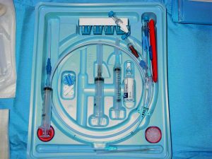
      • Treated according to EGDT protocol (see Figure 2, or below) in ED for at least six hours
        • 500ml bolus of crystalloid q30min to achieve CVP 8-12mm
        • Vasopressors to achieve MAP ≥ 65
        • Vasodilators to achieve MAP ≤ 90
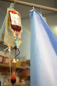
        • If ScvO2 < 70%, transfuse RBCs to achieve Hct ≥ 30
        • If, after CVP, MAP, and Hct were optimized as above and ScvO2 remained < 70%, dobutamine was added and uptitrated to achieve ScvO2 ≥ 70 or until max dose 20 μg/kg/min
          • dobutamine was de-escalated if MAP < 65 or HR > 120
        • Patients in whom hemodynamics could not be optimized were intubated and sedated, in order to decrease oxygen consumption
      • Patients were transferred to inpatient ICU bed as soon as able, and upon transfer ScvO2 measurement was discontinued
      • Inpatient team was blinded to treatment group assignment

The primary outcome was in-hospital mortality. Secondary endpoints included: resuscitation end points, organ-dysfunction scores, coagulation-related variables, administered treatments, and consumption of healthcare resources.

130 patients were randomized to EGDT, and 133 to standard therapy. There were no differences in baseline characteristics. There was no group difference in the prevalence of antibiotics given within the first 6 hours. Standard-therapy patients spent 6.3 ± 3.2 hours in the ED, whereas EGDT patients spent 8.0 ± 2.1 (p < 0.001).

In-hospital mortality was 46.5% in the standard-therapy group, and 30.5% in the EGDT group (p = 0.009, NNT 6.25). 28-day and 60-day mortalities were also improved in the EGDT group. See Table 3.

During the initial six hours of resuscitation, there was no significant group difference in mean heart rate or CVP. MAP was higher in the EGDT group (p < 0.001), but all patients in both groups reached a MAP ≥ 65. ScvO2 ≥ 70% was met by 60.2% of standard-therapy patients and 94.9% of EGDT patients (p < 0.001). A combination endpoint of achievement of CVP, MAP, and UOP (≥ 0.5cc/kg/hr) goals was met by 86.1% of standard-therapy patients and 99.2% of EGDT patients (p < 0.001). Standard-therapy patients had lower ScvO2 and greater base deficit, while lactate and pH values were similar in both groups.

During the period of 7 to 72 hours, the organ-dysfunction scores of APACHE II, SAPS II, and MODS were higher in the standard-therapy group (see Table 2). The prothrombin time, fibrin-split products concentration, and d-dimer concentrations were higher in the standard-therapy group, while PTT, fibrinogen concentration, and platelet counts were similar.

During the initial six hours, EGDT patients received significantly more fluids, pRBCs, and inotropic support than standard-therapy patients. Rates of vasopressor use and mechanical ventilation were similar. During the period of 7 to 72 hours, standard-therapy patients received more fluids, pRBCs, and vasopressors than the EGDT group, and they were more likely to be intubated and to have pulmonary-artery catheterization. Rates of inotrope use were similar. Overall, during the first 72 hrs, standard-therapy patients were more likely to receive vasopressors, be intubated, and undergo pulmonary-artery catheterization. EGDT patients were more likely to receive pRBC transfusion. There was no group difference in total volume of fluid administration or inotrope use. Regarding utilization, there were no group differences in mean duration of vasopressor therapy, mechanical ventilation, or length of stay. Among patients who survived to discharge, standard-therapy patients spent longer in the hospital than EGDT patients (18.4 ± 15.0 vs. 14.6 ± 14.5 days, respectively, p = 0.04).

In conclusion, early goal-directed therapy reduced in-hospital mortality in patients presenting to the ED with severe sepsis or septic shock when compared with usual care. In their discussion, the authors note that “when early therapy is not comprehensive, the progression to severe disease may be well under way at the time of admission to the intensive care unit” (1376).

The Rivers trial has been cited over 11,000 times. It has been widely discussed and dissected for decades. Most importantly, it helped catalyze a then-ongoing paradigm shift of what “usual care” in sepsis is. As noted by our own Drs. Sonti and Vinayak and in their Georgetown Critical Care Top 40: “Though we do not use the ‘Rivers protocol’ as written, concepts (timely resuscitation) have certainly infiltrated our ‘standard of care’ approach.” The Rivers trial evaluated the effect of a bundle (multiple interventions). It was a relatively complex protocol, and it has been recognized that the transfusion of blood to Hgb > 10 may have caused significant harm. In aggregate, the most critical elements of the modern initial resuscitation in sepsis are early administration of antibiotics (notably not protocolized by Rivers) within the first hour and the aggressive administration of IV fluids (now usually 30cc/kg of crystalloid within the first 3 hours of presentation).

More recently, there have been three large RCTs of EGDT versus usual care and/or protocols that used some of the EGDT targets: ProCESS (2014, USA), ARISE (2014, Australia), and ProMISe (2015, UK). In general terms, EGDT provided no mortality benefit compared to usual care. Prospectively, the authors of these three trials planned a meta-analysis – the 2017 PRISM study – which concluded that “EGDT did not result in better outcomes than usual care and was associated with higher hospitalization costs across a broad range of patient and hospital characteristics.” Despite patients in the Rivers trial being sicker than those of ProCESS/ARISE/ProMISe, it was not found in the subgroup analysis of PRISM that EGDT was more beneficial in sicker patients. Overall, the PRISM authors noted that “it remains possible that general advances in the provision of care for sepsis and septic shock, to the benefit of all patients, explain part or all of the difference in findings between the trial by Rivers et al. and the more recent trials.”

Further Reading/References:
1. Rivers trial @ Wiki Journal Club
2. Rivers trial @ 2 Minute Medicine
3. “Early Goal Directed Therapy in Septic Shock” @ Life in The Fast Lane
4. Georgetown Critical Care Top 40
5. “A randomized trial of protocol-based care for early septic shock” (ProCESS). NEJM 2014.
6. “Goal-directed resuscitation for patients with early septic shock” (ARISE). NEJM 2014.
7. “Trial of early, goal-directed resuscitation for septic shock” (ProMISe). NEJM 2015.
8. “Early, Goal-Directed Therapy for Septic Shock – A Patient-level Meta-Analysis” PRISM. NEJM 2017.
9. “Hour-1 Bundle,” Surviving Sepsis Campaign
10. UpToDate, “Evaluation and management of suspected sepsis and septic shock in adults”

Summary by Duncan F. Moore, MD

Image Credit: by Clinical_Cases, CC BY-SA 2.5 , via Wikimedia Commons

Week 4 – ARDSNet

“Ventilation with Lower Tidal Volumes as Compared with Traditional Tidal Volumes for Acute Lung Injury and the Acute Respiratory Distress Syndrome”

by the Acute Respiratory Distress Syndrome Network (ARDSNet)

N Engl J Med. 2000 May 4;342(18):1301-8. [free full text]

Acute respiratory distress syndrome (ARDS) is an inflammatory and highly morbid lung injury found in many critically ill patients. In the 1990s, it was hypothesized that overdistention of aerated lung volumes and elevated airway pressures might contribute to the severity of ARDS, and indeed some work in animal models supported this theory. Prior to the ARDSNet study, four randomized trials had been conducted to investigate the possible protective effect of ventilation with lower tidal volumes, but their results were conflicting.

The ARDSNet study enrolled patients with ARDS (diagnosed within 36 hours) to either a lower initial tidal volume of 6ml/kg, downtitrated as necessary to maintain plateau pressure ≤ 30 cm H2O, or to the “traditional” therapy of an initial tidal volume of 12 ml/kg, downtitrated as necessary to maintain plateau pressure ≤ 50 cm of water. The primary outcomes were in-hospital mortality and ventilator-free days within the first 28 days. Secondary outcomes included number of days without organ failure, occurrence of barotrauma, and reduction in IL-6 concentration from day 0 to day 3.

861 patients were randomized before the trial was stopped early due to the increased mortality in the control arm noted during interim analysis. In-hospital mortality was 31.0% in the lower tidal volume group and 39.8% in the traditional tidal volume group (p = 0.007, NNT = 11.4). Ventilator free days were 12±11 in the lower tidal volume group vs. 10±11 in the traditional group (n = 0.007). The lower tidal volume group had more days without organ failure (15±11 vs. 12±11, p = 0.006). There was no difference in rates of barotrauma among the two groups. Decrease in IL-6 concentration between days 0 and 3 was greater in the low tidal volume group (p < 0.001), and IL-6 concentration at day 3 was lower in the low tidal volume group (p = 0.002).

In summary, low tidal volume ventilation decreases mortality in ARDS relative to “traditional” tidal volumes. The authors felt that this study confirmed the results of prior animal models and conclusively answered the question of whether or not low tidal volume ventilation provided a mortality benefit. In fact, in the years following, low tidal volume ventilation became the standard of care, and a robust body of literature followed this study to further delineate a “lung-protective strategy.” Critics of the study noted that, at the time of the study, the “traditional” (standard of care) tidal volume in ARDS was less than the 12 ml/kg used in the comparison arm. (Non-enrolled patients at the participating centers were receiving a mean tidal volume of 10.3 ml/kg.) Thus not only was the trial making a comparison to a faulty control, but it was also potentially harming patients in the control arm. An excellent summary of the ethical issues and debate regarding this specific issue and regarding control arms of RCTs in general can be found here.

Corresponding practice point from Dr. Sonti and Dr. Vinayak and their Georgetown Critical Care Top 40: “Low tidal volume ventilation is the standard of care in patients with ARDS (P/F < 300). Use ≤ 6 ml/kg predicted body weight, follow plateau pressures, and be cautious of mixed modes in which you set a tidal volume but the ventilator can adjust and choose a larger one.”

PulmCCM is an excellent blog, and they have a nice page reviewing this topic and summarizing some of the research and guidelines that have followed.

Further Reading/References:
1. ARDSNet @ Wiki Journal Club
2. ARDSNet @ 2 Minute Medicine
3. PulmCCM “Mechanical Ventilation in ARDS: Research Update”
4. Georgetown Critical Care Top 40, page 6
5. PulmCCM “In ARDS, substandard ventilator care is the norm, not the exception.” 2017.

Summary by Duncan F. Moore, MD

Week 40 – PROSEVA

Prone Positioning in Severe Acute Respiratory Distress Syndrome
by the PROSEVA Study Group

N Engl J Med. 2013 June 6; 368(23):2159-2168 [free full text]

Prone positioning had been used for many years in ICU patients with ARDS in order to improve oxygenation. Per Dr. Sonti’s Georgetown Critical Care Top 40, the physiologic basis for benefit with proning lies in the idea that atelectatic regions of lung typically occur in the most dependent portion of an ARDS patient, with hyperinflation affecting the remaining lung. Periodic reversal of these regions via moving the patient from supine to prone and vice versa ensures no one region of the lung will have extended exposure to either atelectasis or overdistention. Although the oxygenation benefits have been long noted, the PROSEVA trial established mortality benefit.

Study patients were selected from 26 ICUs in France and 1 in Spain which had daily practice with prone positioning for at least 5 years. Inclusion criteria: ARDS patients intubated and ventilated <36hr with severe ARDS (defined as PaO2:FiO2 ratio <150, PEEP>5, and TV of about 6ml/kg of predicted body weight). (NB: by the Berlin definition for ARDS, severe ARDS is defined as PaO2:FiO2 ratio <100.) Patients were either randomized to the intervention of proning within 36 hours of mechanical ventilation for at least 16 consecutive hours (N=237) or to the control of being left in a semirecumbent (supine) position (N=229). The primary outcome was mortality at day 28. Secondary outcomes included mortality at day 90, rate of successful extubation (no reintubation or use of noninvasive ventilation x48hr), time to successful extubation, length of stay in the ICU, complications, use of noninvasive ventilation, tracheotomy rate, number of days free from organ dysfunction, ventilator settings, measurements of ABG, and respiratory system mechanics during the first week after randomization.

At the time of randomization in the study, the majority of characteristics were similar between the two groups, although the authors noted differences in the SOFA score and the use of neuromuscular blockers and vasopressors. The supine group at baseline had a higher SOFA score indicating more severe organ failure, and also had higher rate of vasopressor usage. The prone group had a higher rate of usage of neuromuscular blockade. The primary outcome of 28 day mortality was significantly lower in the prone group than in the supine group, at 16.0% vs 32.8% (p < 0.001, NNT = 6.0). This mortality decrease was still statistically significant when adjusted for the SOFA score. Secondary outcomes were notable for a significantly higher rate of successful extubation in the prone group (hazard ratio 0.45; 95% CI 0.29-0.7, p < 0.001). Additionally, the PaO2:FiO2 ratio was significantly higher in the supine group, whereas the PEEP and FiO2 were significantly lower. The remainder of secondary outcomes were statistically similar.

PROSEVA showed a significant mortality benefit with early use of prone positioning in severe ARDS. This mortality benefit was considerably larger than that seen in past meta-analyses, which was likely due to this study selecting specifically for patients with severe disease as well as specifying longer prone-positioning sessions than employed in prior studies. Critics have noted the unexpected difference in baseline characteristics between the two arms of the study. While these critiques are reasonable, the authors mitigate at least some of these complaints by adjusting the mortality for the statistically significant differences. With such a radical mortality benefit it might be surprising that more patients are not proned at our institution. One reason is that relatively few of our patients have severe ARDS. Additionally, proning places a high demand on resources and requires a coordinated effort of multiple staff. All treatment centers in this study had specially-trained staff that had been performing proning on a daily basis for at least 5 years, and thus were very familiar with the process. With this in mind, we consider the use of proning in patients meeting criteria for severe ARDS.

References and further reading:
1. PROSEVA @ 2 Minute Medicine
2. PROSEVA @ Wiki Journal Club
3. PROSEVA @ Georgetown Critical Care Top 40, pages 8-9
4. Life in the Fastlane, Critical Care Compendium, “Prone Position and Mechanical Ventilation”
5. PulmCCM.org, “ICU Physiology in 1000 Words: The Hemodynamics of Prone”

Summary by Gordon Pelegrin, MD

Image Credit: by James Heilman, MD, CC BY-SA 3.0, via Wikimedia Commons

Week 35 – CORTICUS

“Hydrocortisone Therapy for Patients with Septic Shock”

N Engl J Med. 2008 Jan 10;358(2):111-24. [free full text]

Steroid therapy in septic shock has been a hotly debated topic since the 1980s. The Annane trial in 2002 suggested that there was a mortality benefit to early steroid therapy and so for almost a decade this became standard of care. In 2008, the CORTICUS trial was performed suggesting otherwise.

The trial enrolled ICU patients with septic shock onset with past 72 hrs (defined as SBP < 90 despite fluids or need for vasopressors and hypoperfusion or organ dysfunction from sepsis). Excluded patients included those with an “underlying disease with a poor prognosis,” life expectancy < 24hrs, immunosuppression, and recent corticosteroid use. Patients were randomized to hydrocortisone 50mg IV q6h x5 days plus taper or to placebo injections q6h x5 days plus taper. The primary outcome was 28-day mortality among patients who did not have a response to ACTH stim test (cortisol rise < 9mcg/dL). Secondary outcomes included 28-day mortality in patients who had a positive response to ACTH stim test, 28-day mortality in all patients, reversal of shock (defined as SBP ≥ 90 for at least 24hrs without vasopressors) in all patients and time to reversal of shock in all patients.

In ACTH non-responders (n = 233), intervention vs. control 28-day mortality was 39.2% vs. 36.1%, respectively (p = 0.69). In ACTH responders (n = 254), intervention vs. control 28-day mortality was 28.8% vs. 28.7% respectively (p = 1.00). Reversal of was shock 84.7%% vs. 76.5% (p = 0.13). Among all patients, intervention vs. control 28-day mortality was 34.3% vs. 31.5% (p = 0.51) and reversal of shock 79.7% vs. 74.2% (p = 0.18). The duration of time to reversal of shock was significantly shorter among patients receiving hydrocortisone (per Kaplan-Meier analysis, p<0.001; see Figure 2) with median time to of reversal 3.3 days vs. 5.8 days (95% CI 5.2 – 6.9).

In conclusion, the CORTICUS trial demonstrated no mortality benefit of steroid therapy in septic shock regardless of a patient’s response to ACTH. Despite the lack of mortality benefit, it demonstrated an earlier resolution of shock with steroids. This lack of mortality benefit sharply contrasted with the previous Annane 2002 study. Several reasons have been posited for this difference including poor powering of the CORTICUS study (which did not reach the desired n = 800), inclusion starting within 72 hrs of septic shock vs. Annane starting within 8 hrs, and the overall sicker nature of Annane patients (who were all mechanically ventilated). Subsequent meta-analyses disagree about the mortality benefit of steroids, but meta-regression analyses suggest benefit among the sickest patients. All studies agree about the improvement in shock reversal. The 2016 Surviving Sepsis Campaign guidelines recommend IV hydrocortisone in septic shock in patients who continue to be hemodynamically unstable despite adequate fluid resuscitation and vasopressor therapy.

Per Drs. Sonti and Vinayak of the GUH MICU (excepted from their excellent Georgetown Critical Care Top 40): “Practically, we use steroids when reaching for a second pressor or if there is multiorgan system dysfunction. Our liver patients may have deficient cortisol production due to inadequate precursor lipid production; use of corticosteroids in these patients represents physiologic replacement rather than adjunct supplement.”

The ANZICS collaborative group published the ADRENAL trial in NEJM in 2018 – which demonstrated that “among patients with septic shock undergoing mechanical ventilation, a continuous infusion of hydrocortisone did not result in lower 90-day mortality than placebo.” The authors did note “a more rapid resolution of shock and a lower incidence of blood transfusion” among patients receiving hydrocortisone. The folks at EmCrit argued [https://emcrit.org/emnerd/cc-nerd-case-relative-insufficiency/] that this was essentially a negative study, and thus in the existing context of CORTICUS, the results of the ADRENAL trial do not change our management of refractory septic shock.

Finally, the 2018 APPROCCHSS trial (also by Annane) evaluated the survival benefit hydrocortisone plus fludocortisone vs. placebo in patients with septic shock and found that this intervention reduced 90-day all-cause mortality. At this time, it is difficult truly discern the added information of this trial given its timeframe, sample size, and severity of underlying illness. See the excellent discussion in the following links: WikiJournal Club, PulmCrit, PulmCCM, and UpToDate.

References / Additional Reading:
1. CORTICUS @ Wiki Journal Club
2. CORTICUS @ Minute Medicine
3. Surviving Sepsis Campaign: International Guidelines for Management of Sepsis and Septic Shock (2016), section “Corticosteroids”
4. Annane trial (2002) full text
5. PulmCCM, “Corticosteroids do help in sepsis: ADRENAL trial”
6. UpToDate, “Glucocorticoid therapy in septic shock”

Post by Gordon Pelegrin, MD

Image Credit: LHcheM, CC BY-SA 3.0, via Wikimedia Commons

Week 29 – PneumA

“Comparison of 8 vs 15 Days of Antibiotic Therapy for Ventilator-Associated Pneumonia in Adults”

JAMA. 2003 November 19;290(19):2588-2598. [free full text]

Ventilator-associated pneumonia (VAP) is a frequent complication of mechanical ventilation and, prior to this study, few trials had addressed the optimal duration of antibiotic therapy in VAP. Thus, patients frequently received 14- to 21-day antibiotic courses. As antibiotic stewardship efforts increased and awareness grew of the association between prolonged antibiotic courses and the development of multidrug resistant (MDR) infections, more data were needed to clarify the optimal VAP treatment duration.

This 2003 trial by the PneumA Trial Group was the first large randomized trial to compare shorter (8-day) versus longer (15-day) treatment courses for VAP.

The noninferiority study, carried out in 51 French ICUs, enrolled intubated patients with clinical suspicion for VAP and randomized them to either 8 or 15 days of antimicrobials. Antimicrobial regimens were chosen by the treating clinician. 401 patients met eligibility criteria. 197 were randomized to the 8-day regimen. 204 patients were randomized to the 15-day regimen. Study participants were blinded to randomization assignment until day 8. Analysis was performed using an intention-to-treat model. The primary outcomes measured were death from any cause at 28 days, antibiotic-free days, and microbiologically documented pulmonary infection recurrence.

Study findings demonstrated a similar 28-day mortality in both groups (18.8% mortality in 8-day group vs. 17.2% in 15-day group, group difference 90% CI -3.7% to 6.9%). The 8-day group did not develop more recurrent infections (28.9% in 8-day group vs. 26.0% in 15-day group, group difference 90% CI -3.2% to 9.1%). The 8-day group did have more antibiotic-free days when measured at the 28-day point (13.1 in 8-day group vs. 8.7 in 15-day group, p<0.001). A subgroup analysis did show that more 8-day-group patients who had an initial infection with lactose-nonfermenting GNRs developed a recurrent pulmonary infection, so noninferiority was not established in this specific subgroup (40.6% recurrent GNR infection in 8-day group vs. 25.4% in 15-day group, group difference 90% CI 3.9% to 26.6%).

Implications/Discussion:
There is no benefit to prolonging VAP treatment to 15 days (except perhaps when Pseudomonas aeruginosa is suspected based on gram stain/culture data). Shorter courses of antibiotics for VAP treatment allow for less antibiotic exposure without increasing rates of recurrent infection or mortality.

The 2016 IDSA guidelines on VAP treatment recommend a 7-day course of antimicrobials for treatment of VAP (as opposed to a longer treatment course such as 8-15 days). These guidelines are based on the IDSA’s own large meta-analysis (of 10 randomized trials, including PneumA, as well as an observational study) which demonstrated that shorter courses of antibiotics (7 days) reduce antibiotic exposure and recurrent pneumonia due to MDR organisms without affecting clinical outcomes, such as mortality. Of note, this 7-day course recommendation also applies to treatment of lactose-nonfermenting GNRs, such as Pseudomonas.

When considering the PneumA trial within the context of the newest IDSA guidelines, we see that we now have over 15 years of evidence supporting the use of shorter VAP treatment courses.

Further Reading/References:
1. 2016 IDSA Guidelines for the Management of HAP/VAP
2. Wiki Journal Club
3. PulmCCM “IDSA Guidelines 2016: HAP, VAP & It’s the End of HCAP as We Know It (And I Feel Fine)”
4. PulmCrit “The siren’s call: Double-coverage for ventilator associated PNA”

Summary by Liz Novick, MD

Image Credit: Joseaperez, CC BY-SA 3.0, via Wikimedia Commons