“Mild Therapeutic Hypothermia to Improve the Neurologic Outcome After Cardiac Arrest”
by the Hypothermia After Cardiac Arrest Study Group
N Engl J Med. 2002 Feb 21;346(8):549-56. [free full text]
—
Neurologic injury after cardiac arrest is a significant source of morbidity and mortality. It is hypothesized that brain reperfusion injury (via the generation of free radicals and other inflammatory mediators) following ischemic time is the primary pathophysiologic basis. Animal models and limited human studies have demonstrated that patients treated with mild hypothermia following cardiac arrest have improved neurologic outcome. The 2002 HACA study sought to evaluate prospectively the utility of therapeutic hypothermia in reducing neurologic sequelae and mortality post-arrest.
Population: European patients who achieve return of spontaneous circulation (ROSC) after presenting to the ED in cardiac arrest
inclusion criteria: witnessed arrest, ventricular fibrillation or non-perfusing ventricular tachycardia as initial rhythm, estimated interval 5 to 15 min from collapse to first resuscitation attempt, no more than 60 min from collapse to ROSC, age 18-75
pertinent exclusion: pt already < 30ºC on admission, comatose state prior to arrest d/t CNS drugs, response to commands following ROSC
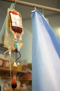
Intervention: Cooling to target temperature 32-34ºC with maintenance for 24 hrs followed by passive rewarming. Pts received pancuronium for neuromuscular blockade to prevent shivering.
Comparison: Standard intensive care
Outcomes:
Primary: a “favorable neurologic outcome” at 6 months defined as Pittsburgh cerebral-performance scale category 1 (good recovery) or 2 (moderate disability). (Of note, the examiner was blinded to treatment group allocation.)
Secondary:
- all-cause mortality at 6 months
- specific complications within the first 7 days: bleeding “of any severity,” pneumonia, sepsis, pancreatitis, renal failure, pulmonary edema, seizures, arrhythmias, and pressure sores
Results:
3551 consecutive patients were assessed for enrollment and ultimately 275 met inclusion criteria and were randomized. The normothermia group had more baseline DM and CAD and were more likely to have received BLS from a bystander prior to the ED.
Regarding neurologic outcome at 6 months, 75 of 136 (55%) of the hypothermia group had a favorable neurologic outcome, versus 54/137 (39%) in the normothermia group (RR 1.40, 95% CI 1.08-1.81, p = 0.009; NNT = 6). After adjusting for all baseline characteristics, the RR increased slightly to 1.47 (95% CI 1.09-1.82).
Regarding death at 6 months, 41% of the hypothermia group had died, versus 55% of the normothermia group (RR 0.74, 95% CI 0.58-0.95, p = 0.02; NNT = 7). After adjusting for all baseline characteristics, RR = 0.62 (95% CI 0.36-0.95). There was no difference among the two groups in the rate of any complication or in the total number of complications during the first 7 days.
Implication/Discussion:
In ED patients with Vfib or pulseless VT arrest who did not have meaningful response to commands after ROSC, immediate therapeutic hypothermia reduced the rate of neurologic sequelae and mortality at 6 months.
Corresponding practice point from Dr. Sonti and Dr. Vinayak and their Georgetown Critical Care Top 40: “If after ROSC your patient remains unresponsive and does not have refractory hypoxemia/hypotension/coagulopathy, you should initiate therapeutic hypothermia even if the arrest was PEA. The benefit seen was substantial and any proposed biologic mechanism would seemingly apply to all causes of cardiac arrest. The investigators used pancuronium to prevent shivering; [at MGUH] there is a ‘shivering’ protocol in place and if refractory, paralytics can be used.”
This trial, as well as a concurrent publication by Benard et al. ushered in a new paradigm of therapeutic hypothermia or “targeted temperature management” (TTM) following cardiac arrest. Numerous trials in related populations and with modified interventions (e.g. target temperature 36º C) were performed over the following decade, and ultimately led to the current standard of practice.
Per UpToDate, the collective trial data suggest that “active control of the post-cardiac arrest patient’s core temperature, with a target between 32 and 36ºC, followed by active avoidance of fever, is the optimal strategy to promote patient survival.” TTM should be undertaken in all patients who do not follow commands or have purposeful movements following ROSC. Expert opinion at UpToDate recommends maintaining temperature control for at least 48 hours.
Further Reading/References:
1. HACA @ 2 Minute Medicine
2. HACA @ Wiki Journal Club
3. HACA @ Visualmed
4. Georgetown Critical Care Top 40, page 23 (Jan. 2016)
5. PulmCCM.org, “Hypothermia did not help after out-of-hospital cardiac arrest, in largest study yet”
6. Cochrane Review, “Hypothermia for neuroprotection in adults after cardiopulmonary resuscitation”
7. The NNT, “Mild Therapeutic Hypothermia for Neuroprotection Following CPR”
8. UpToDate, “Post-cardiac arrest management in adults”
Summary by Duncan F. Moore, MD
