Week 20 – Omeprazole for Bleeding Peptic Ulcers

“Effect of Intravenous Omeprazole on Recurrent Bleeding After Endoscopic Treatment of Bleeding Peptic Ulcers”

N Engl J Med. 2000 Aug 3;343(5):310-6. [free full text]

Intravenous proton-pump inhibitor (PPI) therapy is a cornerstone of modern therapy for bleeding peptic ulcers. However, prior to this 2000 study by Lau et al., the role of PPIs in the prevention of recurrent bleeding after endoscopic treatment was unclear. At the time, re-bleeding rates after endoscopic treatment were noted to be approximately 15-20%. Although other studies had approached this question, no high-quality, large, blinded RCT had examined adjuvant PPI use immediately following endoscopic treatment.

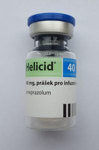
The study enrolled patients who had a bleeding gastroduodenal ulcer visualized on endoscopy and in whom hemostasis was achieved following epinephrine injection and thermocoagulation. Enrollees were randomized to treatment with either omeprazole 80mg IV bolus followed by 8mg/hr infusion x72 hours then followed by omeprazole 20mg PO x8 weeks or to placebo bolus + drip x72 hours followed by omeprazole 20mg PO x8 weeks. The primary outcome was recurrent bleeding within 30 days. Secondary outcomes included recurrent bleeding within 72 hours, amount of blood transfused by day 30, hospitalization duration, and all-cause 30-day mortality.

120 patients were randomized to each arm. The trial was terminated early due to the finding on interim analysis of a significantly lower recurrent bleeding rate in the omeprazole arm. Bleeding re-occurred within 30 days in 8 (6.7%) omeprazole patients versus 27 (22.5%) placebo patients (HR 3.9, 95% CI 1.7-9.0; NNT 6.3). A Cox proportional-hazards model, when adjusted for size and location of ulcers, presence/absence of coexisting illness, and history of ulcer disease, revealed a similar hazard ratio (HR 3.9, 95% CI 1.7-9.1). Recurrent bleeding was most common during the first 72 hrs (4.2% of the omeprazole group versus 20% of the placebo group, RR 4.80, 95% CI 1.89-12.2, p<0.001). For a nice visualization of the early separation of re-bleeding rates, see the Kaplan-Meier curve in Figure 1. The mean number of units of blood transfused within 30 days was 2.7 ± 2.5 in the omeprazole group versus 3.5 ± 3.8 in the placebo group (p = 0.04). Regarding duration of hospitalization, 46.7% of omeprazole patients were admitted for < 5 days versus 31.7% of placebo patients (p = 0.02). Median stay was 4 days in the omeprazole group versus 5 days in the placebo group (p = 0.006). 4.2% of the omeprazole patients died within 30 days, whereas 10% of the placebo patients died (p = 0.13).

Treatment with intravenous omeprazole immediately following endoscopic intervention for bleeding peptic ulcer significantly reduced the rate of recurrent bleeding. This effect was most prominent within the first 3 days of therapy. This intervention also reduced blood transfusion requirements and shortened hospital stays. The presumed mechanism of action is increased gastric pH facilitating platelet aggregation. In 2018, the benefit of this intervention seems so obvious based on its description alone that one would not imagine that such a trial would be funded or published in such a high-profile journal. However, the annals of medicine are littered with now-discarded interventions that made sense from a theoretical or mechanistic perspective but were demonstrated to be ineffective or even harmful (e.g. pharmacologic suppression of ventricular arrhythmias post-MI or renal denervation for refractory HTN).

Today, bleeding peptic ulcers are treated with an IV PPI twice daily. Per UpToDate, meta-analyses have not shown a benefit of continuous PPI infusion over this IV BID dosing. However, per 2012 guidelines in the American Journal of Gastroenterology, patients with active bleeding or non-bleeding visible vessels should receive both endoscopic intervention and IV PPI bolus followed by infusion.

Further Reading/References:
1. Wiki Journal Club
2. 2 Minute Medicine
3. UpToDate, “Overview of the Treatment of Bleeding Peptic Ulcers”
4. Laine L, Jensen DM. “Management of patients with ulcer bleeding.” Am J Gastroenterol. 2012

Summary by Duncan F. Moore, MD

Image credit: Wesalius, CC BY 4.0, via Wikimedia Commons

Week 13 – Sepsis-3

“The Third International Consensus Definitions for Sepsis and Septic Shock (Sepsis-3)”

JAMA. 2016 Feb 23;315(8):801-10. [free full text]

In practice, we recognize sepsis as a potentially life-threatening condition that arises secondary to infection. Because the SIRS criteria were of limited sensitivity and specificity in identifying sepsis and because our understanding of the pathophysiology of sepsis had purportedly advanced significantly during the interval since the last sepsis definition, an international task force of 19 experts was convened to define and prognosticate sepsis more effectively. The resulting 2016 Sepsis-3 definition was the subject of immediate and sustained controversy.

In the words of Sepsis-3, sepsis simply “is defined as life-threatening organ dysfunction caused by a dysregulated host response to infection.” The paper further defines organ dysfunction in terms of a threshold change in the SOFA score by 2+ points. However, the authors state that “the SOFA score is not intended to be used as a tool for patient management but as a means to clinically characterize a septic patient.” The authors note that qSOFA, an easier tool introduced in this paper, can identify promptly at the bedside patients “with suspected infection who are likely to have a prolonged ICU stay or die in the hospital.” A positive screen on qSOFA is identified as 2+ of the following: AMS, SBP ≤ 100, or respiratory rate ≥ 22. At the time of this endorsement of qSOFA, the tool had not been validated prospectively. Finally, septic shock was defined as sepsis with persistent hypotension requiring vasopressors to maintain MAP ≥ 65 and with a serum lactate > 2 despite adequate volume resuscitation.

As noted contemporaneously in the excellent PulmCrit blog post “Top ten problems with the new sepsis definition,” Sepsis-3 was not endorsed by the American College of Chest Physicians, the IDSA, any emergency medicine society, or any hospital medicine society. On behalf of the American College of Chest Physicians, Dr. Simpson published a scathing rejection of Sepsis-3 in Chest in May 2016. He noted “there is still no known precise pathophysiological feature that defines sepsis.” He went on to state “it is not clear to us that readjusting the sepsis criteria to be more specific for mortality is an exercise that benefits patients,” and said “to abandon one system of recognizing sepsis [SIRS] because it is imperfect and not yet in universal use for another system that is used even less seems unwise without prospective validation of that new system’s utility.”

In fact, the later validation of qSOFA demonstrated that the SIRS criteria had superior sensitivity for predicting in-hospital mortality while qSOFA had higher specificity. See the following posts at PulmCrit for further discussion: [https://emcrit.org/isepsis/isepsis-sepsis-3-0-much-nothing/] [https://emcrit.org/isepsis/isepsis-sepsis-3-0-flogging-dead-horse/].

At UpToDate, authors note that “data of the value of qSOFA is conflicting,” and because of this, “we believe that further studies that demonstrate improved clinically meaningful outcomes due to the use of qSOFA compared to clinical judgement are warranted before it can be routinely used to predict those at risk of death from sepsis.”

Additional Reading:
1. PulmCCM, “Simple qSOFA score predicts sepsis as well as anything else”
2. 2 Minute Medicine

Summary by Duncan F. Moore, MD

Image Credit: By Mark Oniffrey – Own work, CC BY-SA 4.0

Week 12 – Rivers Trial

“Early Goal-Directed Therapy in the Treatment of Severe Sepsis and Septic Shock”

N Engl J Med. 2001 Nov 8;345(19):1368-77. [free full text]

Sepsis is common and, in its more severe manifestations, confers a high mortality risk. Fundamentally, sepsis is a global mismatch between oxygen demand and delivery. Around the time of this seminal study by Rivers et al., there was increasing recognition of the concept of the “golden hour” in sepsis management – “where definitive recognition and treatment provide maximal benefit in terms of outcome” (1368). Rivers and his team created a “bundle” of early sepsis interventions that targeted preload, afterload, and contractility, dubbed early goal-directed therapy (EGDT). They evaluated this bundle’s effect on mortality and end-organ dysfunction.

The “Rivers trial” randomized adults presenting to a single US academic center ED with ≥ 2 SIRS criteria and either SBP ≤ 90 after a crystalloid challenge of 20-30ml/kg over 30min or lactate > 4mmol/L to either treatment with the EGDT bundle or to the standard of care.

Intervention: early goal-directed therapy (EGDT)

  • Received a central venous catheter with continuous central venous O2 saturation (ScvO2) measurement
  • Treated according to EGDT protocol (see Figure 2, or below) in ED for at least six hours
    • 500ml bolus of crystalloid q30min to achieve CVP 8-12mm
    • Vasopressors to achieve MAP ≥ 65
    • Vasodilators to achieve MAP ≤ 90
    • If ScvO2 < 70%, transfuse RBCs to achieve Hct ≥ 30
    • If, after CVP, MAP, and Hct were optimized as above and ScvO2 remained < 70%, dobutamine was added and uptitrated to achieve ScvO2 ≥ 70 or until max dose 20 μg/kg/min
      • dobutamine was de-escalated if MAP < 65 or HR > 120
    • Patients in whom hemodynamics could not be optimized were intubated and sedated, in order to decrease oxygen consumption
  • Patients were transferred to inpatient ICU bed as soon as able, and upon transfer ScvO2 measurement was discontinued
  • Inpatient team was blinded to treatment group assignment

The primary outcome was in-hospital mortality. Secondary endpoints included: resuscitation end points, organ-dysfunction scores, coagulation-related variables, administered treatments, and consumption of healthcare resources.

130 patients were randomized to EGDT, and 133 to standard therapy. There were no differences in baseline characteristics. There was no group difference in the prevalence of antibiotics given within the first 6 hours. Standard-therapy patients spent 6.3 ± 3.2 hours in the ED, whereas EGDT patients spent 8.0 ± 2.1 (p < 0.001).

In-hospital mortality was 46.5% in the standard-therapy group, and 30.5% in the EGDT group (p = 0.009, NNT 6.25). 28-day and 60-day mortalities were also improved in the EGDT group. See Table 3.

During the initial six hours of resuscitation, there was no significant group difference in mean heart rate or CVP. MAP was higher in the EGDT group (p < 0.001), but all patients in both groups reached a MAP ≥ 65. ScvO2 ≥ 70% was met by 60.2% of standard-therapy patients and 94.9% of EGDT patients (p < 0.001). A combination endpoint of achievement of CVP, MAP, and UOP (≥ 0.5cc/kg/hr) goals was met by 86.1% of standard-therapy patients and 99.2% of EGDT patients (p < 0.001). Standard-therapy patients had lower ScvO2 and greater base deficit, while lactate and pH values were similar in both groups.

During the period of 7 to 72 hours, the organ-dysfunction scores of APACHE II, SAPS II, and MODS were higher in the standard-therapy group (see Table 2). The prothrombin time, fibrin-split products concentration, and d-dimer concentrations were higher in the standard-therapy group, while PTT, fibrinogen concentration, and platelet counts were similar.

During the initial six hours, EGDT patients received significantly more fluids, pRBCs, and inotropic support than standard-therapy patients. Rates of vasopressor use and mechanical ventilation were similar. During the period of 7 to 72 hours, standard-therapy patients received more fluids, pRBCs, and vasopressors than the EGDT group, and they were more likely to be intubated and to have pulmonary-artery catheterization. Rates of inotrope use were similar. Overall, during the first 72 hrs, standard-therapy patients were more likely to receive vasopressors, be intubated, and undergo pulmonary-artery catheterization. EGDT patients were more likely to receive pRBC transfusion. There was no group difference in total volume of fluid administration or inotrope use. Regarding utilization, there were no group differences in mean duration of vasopressor therapy, mechanical ventilation, or length of stay. Among patients who survived to discharge, standard-therapy patients spent longer in the hospital than EGDT patients (18.4 ± 15.0 vs. 14.6 ± 14.5 days, respectively, p = 0.04).

In conclusion, early goal-directed therapy reduced in-hospital mortality in patients presenting to the ED with severe sepsis or septic shock when compared with usual care. In their discussion, the authors note that “when early therapy is not comprehensive, the progression to severe disease may be well under way at the time of admission to the intensive care unit” (1376).

The Rivers trial has been cited over 10,500 times. It has been widely discussed and dissected for decades. Most importantly, it helped catalyze a then-ongoing paradigm shift of what “usual care” in sepsis is. As noted by our own Drs. Sonti and Vinayak and in their Georgetown Critical Care Top 40: “Though we do not use the ‘Rivers protocol’ as written, concepts (timely resuscitation) have certainly infiltrated our ‘standard of care’ approach.” The Rivers trial evaluated the effect of a bundle (multiple interventions). It was a relatively complex protocol, and it has been recognized that the transfusion of blood to Hgb > 10 may have caused significant harm. In aggregate, the most critical elements of the modern initial resuscitation in sepsis are early administration of antibiotics (notably not protocolized by Rivers) within the first hour and the aggressive administration of IV fluids (now usually 30cc/kg of crystalloid within the first 3 hours of presentation).

More recently, there have been three large RCTs of EGDT versus usual care and/or protocols that used some of the EGDT targets: ProCESS (2014, USA), ARISE (2014, Australia), and ProMISe (2015, UK). In general terms, EGDT provided no mortality benefit compared to usual care. Prospectively, the authors of these three trials planned a meta-analysis – the 2017 PRISM study – which concluded that “EGDT did not result in better outcomes than usual care and was associated with higher hospitalization costs across a broad range of patient and hospital characteristics.” Despite patients in the Rivers trial being sicker than those of ProCESS/ARISE/ProMISe, it was not found in the subgroup analysis of PRISM that EGDT was more beneficial in sicker patients. Overall, the PRISM authors noted that “it remains possible that general advances in the provision of care for sepsis and septic shock, to the benefit of all patients, explain part or all of the difference in findings between the trial by Rivers et al. and the more recent trials.”

Further Reading/References:
1. Wiki Journal Club
2. 2 Minute Medicine
3. Life in The Fast Lane
4. Georgetown Critical Care Top 40
5. “A randomized trial of protocol-based care for early septic shock” (ProCESS). NEJM 2014.
6. “Goal-directed resuscitation for patients with early septic shock” (ARISE). NEJM 2014.
7. “Trial of early, goal-directed resuscitation for septic shock” (ProMISe). NEJM 2015.
8. “Early, Goal-Directed Therapy for Septic Shock – A Patient-level Meta-Analysis” PRISM. NEJM 2017.
9. Surviving Sepsis Campaign
10. UpToDate, “Evaluation and management of suspected sepsis and septic shock in adults”

Summary by Duncan F. Moore, MD

Image Credit: By Clinical_Cases, [CC BY-SA 2.5] via Wikimedia Commons

Week 9 – NICE-SUGAR

“Intensive versus Conventional Glucose Control in Critically Ill Patients”

by the Normoglycemia in Intensive Care Evaluation–Survival Using Glucose Algorithm Regulation (NICE-SUGAR) investigators

N Engl J Med 2009;360:1283-97. [free full text]

On the wards we often hear 180 mg/dL used as the upper limit of acceptable for blood glucose with the understanding that tighter glucose control in inpatients can lead to more harm than benefit. The relevant evidence base comes from ICU populations, with scant direct data in non-ICU patients. The 2009 NICE-SUGAR study is the largest and best among this evidence base.

The study randomized ICU patients (expected to require 3 or more days of ICU-level care) to either “intensive” glucose control (target glucose 81 to 108 mg/dL) or conventional glucose control (target of less than 180 mg/dL). The primary outcome was 90-day all-cause mortality.

6104 patients were randomized to the two arms, and both groups had similar baseline characteristics. 27.5% of patients in the intensive-control group died versus 24.9% in the conventional-control group (OR 1.14, 95% CI 1.02-1.28, p= 0.02). Severe hypoglycemia (< 40 mg/dL) was found in 6.8% of intensive patients but only 0.5% of conventional patients.

In conclusion, intensive glucose control increases mortality in ICU patients. The fact that only 20% of these patients had diabetes mellitus suggests that much of the hyperglycemia treated in this study (97% of intensive group received insulin, 69% of conventional) was from stress, critical illness, and corticosteroid use. For ICU patients, intensive insulin therapy is clearly harmful, but the ideal target glucose range remains controversial and by expert opinion appears to be 140-180. For non-ICU inpatients with or without diabetes mellitus, the ideal glucose target is also unclear – the ADA recommends 140-180, and the Endocrine Society recommends a pre-meal target of < 140 and random levels < 180.

References / Further Reading:
1. ADA Standards of Medical Care in Diabetes 2016 (skip to page S99)
2. Wiki Journal Club
3. Visual Abstract @ VisualMed

Summary by Duncan F. Moore, MD

Image Credit: Dietmar Rabich / Wikimedia Commons / “Würfelzucker — 2018 — 3564” / CC BY-SA 4.0

Week 7 – ARDSNet aka ARMA

“Ventilation with Lower Tidal Volumes as Compared with Traditional Tidal Volumes for Acute Lung Injury and the Acute Respiratory Distress Syndrome”

by the Acute Respiratory Distress Syndrome Network (ARDSNet)

N Engl J Med. 2000 May 4;342(18):1301-8. [free full text]


Acute respiratory distress syndrome (ARDS) is an inflammatory and highly morbid lung injury found in many critically ill patients. In the 1990s, it was hypothesized that overdistention of aerated lung volumes and elevated airway pressures might contribute to the severity of ARDS, and indeed some work in animal models supported this theory. Prior to the ARDSNet study, four randomized trials had been conducted to investigate the possible protective effect of ventilation with lower tidal volumes, but their results were conflicting.

The ARDSNet study enrolled patients with ARDS (diagnosed within 36 hours) to either a lower initial tidal volume of 6ml/kg, downtitrated as necessary to maintain plateau pressure ≤ 30 cm H2O, or to the “traditional” therapy of an initial tidal volume of 12 ml/kg, downtitrated as necessary to maintain plateau pressure ≤ 50 cm of water. The primary outcomes were in-hospital mortality and ventilator-free days within the first 28 days. Secondary outcomes included number of days without organ failure, occurrence of barotrauma, and reduction in IL-6 concentration from day 0 to day 3.

861 patients were randomized before the trial was stopped early due to the increased mortality in the control arm noted during interim analysis. In-hospital mortality was 31.0% in the lower tidal volume group and 39.8% in the traditional tidal volume group (p = 0.007, NNT = 11.4). Ventilator free days were 12±11 in the lower tidal volume group vs. 10±11 in the traditional group (n = 0.007). The lower tidal volume group had more days without organ failure (15±11 vs. 12±11, p = 0.006). There was no difference in rates of barotrauma among the two groups. Decrease in IL-6 concentration between days 0 and 3 was greater in the low tidal volume group (p < 0.001), and IL-6 concentration at day 3 was lower in the low tidal volume group (p = 0.002).

In summary, low tidal volume ventilation decreases mortality in ARDS relative to “traditional” tidal volumes. The authors felt that this study confirmed the results of prior animal models and conclusively answered the question of whether or not low tidal volume ventilation provided a mortality benefit. In fact, in the years following, low tidal volume ventilation became the standard of care, and a robust body of literature followed this study to further delineate a “lung-protective strategy.” Critics of the study noted that, at the time of the study, the “traditional” (standard of care) tidal volume in ARDS was less than the 12 ml/kg used in the comparison arm. (Non-enrolled patients at the participating centers were receiving a mean tidal volume of 10.3 ml/kg.) Thus not only was the trial making a comparison to a faulty control, but it was also potentially harming patients in the control arm. An excellent summary of the ethical issues and debate regarding this specific issue and regarding control arms of RCTs in general can be found here.

Corresponding practice point from Dr. Sonti and Dr. Vinayak and their Georgetown Critical Care Top 40: “Low tidal volume ventilation is the standard of care in patients with ARDS (P/F < 300). Use ≤ 6 ml/kg predicted body weight, follow plateau pressures, and be cautious of mixed modes in which you set a tidal volume but the ventilator can adjust and choose a larger one.”

PulmCCM is an excellent blog, and they have a nice page reviewing this topic and summarizing some of the research and guidelines that have followed.

Further Reading/References:
1. Wiki Journal Club
2. 2 Minute Medicine
3. PulmCCM “Mechanical Ventilation in ARDS: Research Update”
4. Georgetown Critical Care Top 40, page 6
5. PulmCCM “In ARDS, substandard ventilator care is the norm, not the exception.” 2017.

Summary by Duncan F. Moore, MD

Photo Credit: Hanno H. Endres at de.wikipedia, CC BY-SA 3.0

Week 4 – Dexamethasone in Bacterial Meningitis

Streptococcus pneumoniae

“Dexamethasone in Adults With Bacterial Meningitis”

N Engl J Med 2002; 347:1549-1556. [free full text]

The current standard of care in the treatment of suspected bacterial meningitis in the developed world includes the administration of dexamethasone prior to or at the time of antibiotic initiation. The initial evaluation of this practice in part stemmed from animal studies, which demonstrated that dexamethasone reduced CSF concentrations of inflammatory markers as well as neurologic sequelae after meningitis. RCTs in the pediatric literature also demonstrated clinical benefit. The best prospective trial in adults was this 2002 study by de Gans et al.

The trial enrolled adults with suspected meningitis and randomized them to either dexamethasone 10mg IV q6hrs x4 days started 15-20 minutes before the first IV antibiotics or a placebo IV with the same administration schedule. The primary outcome was the Glasgow Outcome Scale at 8 weeks (1 = death, 2 = vegetative state, 3 = unable to live independently, 4 = unable to return to school/work, 5 = able to return to school/work). Secondary outcomes included death and focal neurologic abnormalities. Subgroup analyses were performed by organism.

301 patients were randomized. At 8 weeks, 15% of dexamethasone patients compared with 25% of placebo patients had an unfavorable outcome of Glasgow Outcome Scale score 1-4 (RR 0.59, 95% CI 0.37 – 0.94, p= 0.03). Among patients with pneumococcal meningitis, 26% of dexamethasone patients compared with 52% of placebo patients had an unfavorable outcome. There was no significant difference among treatment arms within the subgroup of patients infected with meningococcal meningitis. Overall, death occurred in 7% of dexamethasone patients and 15% of placebo patients (RR 0.48, 95% CI 0.24 – 0.96, p = 0.04). In pneumococcal meningitis, 14% of dexamethasone patients died, and 34% of placebo patients died.  There was no difference in rates of focal neurologic abnormalities or hearing loss in either treatment arm (including within any subgroup).

In conclusion, early adjunctive dexamethasone improves mortality in bacterial meningitis. As noted in the above subgroup analysis, this benefit appears to be driven by the efficacy within the pneumococcal meningitis subgroup. Of note, the standard initial treatment regimen in this study was amoxicillin 2gm q4hrs for 7-10 days rather than our standard ceftriaxone + vancomycin +/- ampicillin. Largely on the basis of this study alone, the IDSA guidelines for the treatment of bacterial meningitis (2004) recommend dexamethasone 0.15 mg/kg q6hrs for 2-4 days with first dose administered 10-20 min before or concomitant with initiation of antibiotics. Dexamethasone should be continued only if CSF Gram stain, CSF culture, or blood cultures are consistent with pneumococcus.

References / Further Reading:
1. IDSA guidelines for management of bacterial meningitis (2004)
2. Wiki Journal Club
3. 2 Minute Medicine

Summary by Duncan F. Moore, MD

Photo Credit: CDC/Janice Carr. Content Providers(s): CDC/Dr. Richard Facklam. Public Health Image Library #262.

Week 2 – Albumin in SBP

“Effect of Intravenous Albumin on Renal Impairment and Mortality in Patients with Cirrhosis and Spontaneous Bacterial Peritonitis”

N Engl J Med. 1999 Aug 5;341(6):403-9. [free full text]

Renal failure commonly develops in the setting of SBP, and its development is a sensitive predictor of in-hospital mortality. The renal impairment is thought to stem from decreased effective arterial blood volume secondary to the systemic inflammatory response to the infection. In our current practice, there are certain circumstances in which we administer albumin early in the SBP disease course in order to reduce the risk of renal failure and mortality. Ultimately, our current protocol originated from the 1999 study of albumin in SBP by Sort et al.

The trial enrolled adults with SBP and randomized them to treatment with either cefotaxime and albumin infusion 1.5 gm/kg within 6hrs of enrollment, followed by 1 gm/kg on day 3 or cefotaxime alone. The primary outcome was the development of “renal impairment” (a “nonreversible” increase in BUN or Cr by more than 50% to a value greater than 30 mg/dL or 1.5 mg/dL, respectively) during hospitalization. The secondary outcome was in-hospital mortality.

126 patients were randomized. Both groups had similar baseline characteristics, and both had similar rates of resolution of infection. Renal impairment occurred in 10% of the albumin group and 33% of the cefotaxime-alone group (p=0.02). In-hospital mortality was 10% in the albumin group and 29% in the cefotaxime-alone group (p=0.01). 78% of patients that developed renal impairment died in-hospital, while only 3% of patients who did not develop renal impairment died. Plasma renin activity was significantly higher on days 3, 6, and 9 in the cefotaxime-alone group than in the albumin group, while there were no significant differences in MAP among the two groups at those time intervals. Multivariate analysis of all trial participants revealed that baseline serum bilirubin and creatinine were independent predictors of the development of renal impairment.

In conclusion, albumin administration reduces renal impairment and improves mortality in patients with SBP. The findings of this landmark trial were refined by a brief 2007 report by Sigal et al. entitled “Restricted use of albumin for spontaneous bacterial peritonitis.” “High-risk” patients, identified by baseline serum bilirubin of ≥ 4.0 mg/dL or Cr ≥ 1.0 mg/dL were given the intervention of albumin 1.5gm/kg on day 1 and 1gm/kg on day 3, and low-risk patients were not given albumin. None of the 15 low-risk patients developed renal impairment or died, whereas 12 of 21 (57%) of the high-risk group developed renal impairment, and 5 of the 21 (24%) died. The authors conclude that patients with bilirubin < 4.0 and Cr < 1.0 did not need scheduled albumin in the treatment of SBP. The current (2012) American Association for the Study of Liver Diseases guidelines for the management of adult patients with ascites due to cirrhosis do not definitively recommend criteria for albumin administration in SBP. Instead they summarize the aforementioned two studies. A 2013 meta-analysis of four reports/trials (including the two above) concluded that albumin infusion reduced renal impairment and improved mortality with pooled odds ratios approximately commensurate with those of the 1999 study by Sort et al. Ultimately, the current recommended practice per expert opinion is to perform albumin administration per the protocol outlined by Sigal et al. (2007).

References / Further Reading:
1. AASLD Guidelines for Management of Adult Patients with Ascites Due to Cirrhosis (skip to page 77)
2. Sigal et al. “Restricted use of albumin for spontaneous bacterial peritonitis.” Gut 2007.
3. Meta-analysis: “Albumin infusion improves outcomes of patients with spontaneous bacterial peritonitis: a meta-analysis of randomized trials”
4. Wiki Journal Club
5. 2 Minute Medicine

Summary by Duncan F. Moore, MD

Week 50 – Sepsis-3

“The Third International Consensus Definitions for Sepsis and Septic Shock (Sepsis-3)”

JAMA. 2016 Feb 23;315(8):801-10. [free full text]

In practice, we recognize sepsis as a potentially life-threatening condition that arises secondary to infection.  Because the SIRS criteria were of limited sensitivity and specificity in identifying sepsis and because our understanding of the pathophysiology of sepsis had purportedly advanced significantly during the interval since the last sepsis definition, an international task force of 19 experts was convened to define and prognosticate sepsis more effectively. The resulting 2016 Sepsis-3 definition was the subject of immediate and sustained controversy.

In the words of Sepsis-3, sepsis simply “is defined as life-threatening organ dysfunction caused by a dysregulated host response to infection.” The paper further defines organ dysfunction in terms of a threshold change in the SOFA score by 2+ points. However, the authors state that “the SOFA score is not intended to be used as a tool for patient management but as a means to clinically characterize a septic patient.” The authors note that qSOFA, an easier tool introduced in this paper, can identify promptly at the bedside patients “with suspected infection who are likely to have a prolonged ICU stay or die in the hospital.” A positive screen on qSOFA is identified as 2+ of the following: AMS, SBP ≤ 100, or respiratory rate ≥ 22. At the time of this endorsement of qSOFA, the tool had not been validated prospectively. Finally, septic shock was defined as sepsis with persistent hypotension requiring vasopressors to maintain MAP ≥ 65 and with a serum lactate > 2 despite adequate volume resuscitation.

As noted contemporaneously in the excellent PulmCrit blog post “Top ten problems with the new sepsis definition,” Sepsis-3 was not endorsed by the American College of Chest Physicians, the IDSA, any emergency medicine society, or any hospital medicine society. On behalf of the American College of Chest Physicians, Dr. Simpson published a scathing rejection of Sepsis-3 in Chest in May 2016. He noted “there is still no known precise pathophysiological feature that defines sepsis.” He went on to state “it is not clear to us that readjusting the sepsis criteria to be more specific for mortality is an exercise that benefits patients,” and said “to abandon one system of recognizing sepsis [SIRS] because it is imperfect and not yet in universal use for another system that is used even less seems unwise without prospective validation of that new system’s utility.”

In fact, the later validation of qSOFA demonstrated that the SIRS criteria had superior sensitivity for predicting in-hospital mortality while qSOFA had higher specificity. See the following posts at PumCrit for further discussion: [https://emcrit.org/isepsis/isepsis-sepsis-3-0-much-nothing/] [https://emcrit.org/isepsis/isepsis-sepsis-3-0-flogging-dead-horse/].

At UpToDate, authors note that “data of the value of qSOFA is conflicting,” and because of this, “we believe that further studies that demonstrate improved clinically meaningful outcomes due to the use of qSOFA compared to clinical judgement are warranted before it can be routinely used to predict those at risk of death from sepsis.”

Additional Reading:
1. PulmCCM, “Simple qSOFA score predicts sepsis as well as anything else”
2. 2 Minute Medicine

Summary by Duncan F. Moore, MD

Week 46 – Transfusion Strategies for Upper GI Bleeding

“Transfusion Strategies for Acute Upper Gastrointestinal Bleeding”

N Engl J Med. 2013 Jan 3;368(1):11-21. [free full text]

A restrictive transfusion strategy of 7 gm/dL was established following the previously discussed 1999 TRICC trial. Notably, both TRICC and its derivative study TRISS excluded patients who had an active bleed. In 2013, Villanueva et al. performed a study to establish whether there was benefit to a restrictive transfusion strategy in patients with acute upper GI bleeding.

Population: adults with hematemesis (or bloody nasogastric aspirate), melena, or both; selected consecutively at a single-center in Spain

Notable exclusion criteria: a clinical Rockall score* of 0 with a hemoglobin level higher than 12g/dL, massive exsanguinating bleeding, lower GIB, patient refusal of blood transfusion, ACS, stroke/TIA, transfusion within 90 days, recent trauma or surgery

*The Rockall score is a system to assess risk for further bleeding or death on a scale from 0-11. Higher scores (3-11) indicate higher risk. Of the 648 patients excluded, the most common reason for exclusion (n = 329) was low risk of bleeding.

Intervention: restrictive transfusion strategy (transfusion threshold Hgb = 7.0 gm/dL) [n = 444]

Comparison: liberal transfusion strategy (transfusion threshold Hgb = 9.0 gm/dL) [n = 445]

During randomization, patients were stratified by presence or absence of cirrhosis.

As part of the study design, all patients underwent emergent EGD within 6 hours and received relevant hemostatic intervention depending on the cause of bleeding.

Outcome:
Primary outcome: 45-day mortality

Secondary outcomes, selected:

  • Incidence of further bleeding associated with hemodynamic instability or hemoglobin drop > 2 gm/dL in 6 hours
  • Incidence and number of RBC transfusions
  • Other products and fluids transfused
  • Hgb level at nadir, discharge, and 45 days

Subgroup analyses: Patients were stratified by presence of cirrhosis and corresponding Child-Pugh class, variceal bleeding, and peptic ulcer bleeding. An additional subgroup analysis was performed to evaluate changes in hepatic venous pressure gradient between the two strategies.


Results
:
The primary outcome of 45-day mortality was lower in the restrictive strategy (5% vs. 9%; HR 0.55, 95% CI 0.33-0.92; p = 0.02; NNT = 24.8). In subgroup analysis, this finding remained consistent for patients who had Child-Pugh class A or B but was not statistically significant among patients who had Class C. Further stratification for variceal bleeding and peptic ulcer disease did not make a difference in mortality.

Secondary outcomes:
Rates of further bleeding events and RBC transfusion, as well as number of products transfused, were lower in the restrictive strategy. Subgroup analysis demonstrated that rates of re-bleeding were lower in Child-Pugh class A and B but not in C. As expected, the restrictive strategy also resulted in the lowest hemoglobin levels at 24 hours. Hemoglobin levels among patients in the restrictive strategy were lower at discharge but were not significantly different from the liberal strategy at 45 days. There was no group difference in amount of non-RBC blood products or colloid/crystalloid transfused. Patients in the restrictive strategy experienced fewer adverse events, particularly transfusion reactions such as transfusion-associated circulatory overload and cardiac complications. Patients in the liberal-transfusion group had significant post-transfusion increases in mean hepatic venous pressure gradient following transfusion. Such increases were not seen in the restrictive-strategy patients.


Implication/Discussion
:
In patients with acute upper GI bleeds, a restrictive strategy with a transfusion threshold 7 gm/dL reduces 45-day mortality, the rate and frequency of transfusions, and the rate of adverse reactions, relative to a liberal strategy with a transfusion threshold of 9 gm/dL.

In their discussion, the authors hypothesize that the “harmful effects of transfusion may be related to an impairment of hemostasis. Transfusion may counteract the splanchnic vascoconstrictive response caused by hypovolemia, inducing an increase in splanchnic blood flow and pressure that may impair the formation of clots. Transfusion may also induce abnormalities in coagulation properties.”

Subgroup analysis suggests that the benefit of the restrictive strategy is less pronounced in patients with more severe hepatic dysfunction. These findings align with prior studies in transfusion thresholds for critically ill patients. However, the authors note that the results conflict with studies in other clinical circumstances, specifically in the pediatric ICU and in hip surgery for high-risk patients.

There are several limitations to this study. First, its exclusion criteria limit its generalizability. Excluding patients with massive exsanguination is understandable given lack of clinical equipoise; however, this choice allows too much discretion with respect to the definition of a massive bleed. (Note that those excluded due to exsanguination comprised only 39 of 648.) Lack of blinding was a second limitation. Potential bias was mitigated by well-defined transfusion protocols. Additionally, there a higher incidence of transfusion-protocol violations in the restrictive group, which probably biased results toward the null. Overall, deviations from the protocol occurred in fewer than 10% of cases.


Further Reading/References
:
1. Transfusion Strategies for Acute Upper GI Bleeding @ Wiki Journal Club
2. 2 Minute Medicine
3. TRISS @ Wiki Journal Club

Summary by Gordon Pelegrin, MD

Week 44 – Early TIPS in Cirrhosis with Variceal Bleeding

“Early Use of TIPS in Patients with Cirrhosis and Variceal Bleeding”

N Engl J Med. 2010 Jun 24;362(25):2370-9. [free full text]

Variceal bleeding is a major cause of morbidity and mortality in decompensated cirrhosis. The standard of care for an acute variceal bleed includes a combination of vasoactive drugs, prophylactic antibiotics, and endoscopic techniques (e.g. banding). Transjugular intrahepatic portosystemic shunt (TIPS) can be used to treat refractory bleeding. This 2010 trial sought to determine the utility of early TIPS during the initial bleed in high-risk patients, when compared to standard therapy.

Population: cirrhotic patients with acute esophageal variceal bleeding, either Child-Pugh class C with score 10-13 or class B (score 7-9) with active bleeding at diagnostic endoscopy

Notable exclusion criteria: Child-Pugh score > 13, age > 75, HCC that did not meet transplantation criteria, bleeding gastric varices, total portal vein thrombosis, prior TIPS

All patients received endoscopic band ligation (EBL) or endoscopic injection sclerotherapy (EIS) at the time of diagnostic endoscopy. All patients also received vasoactive drugs (terlipressin, somatostatin, or octreotide).

Intervention: TIPS performed within 72 hours after diagnostic endoscopy

Comparison: 1) treatment with vasoactive drugs with transition to nonselective beta blocker when patients free of bleeding followed by 2) addition of isosorbide mononitrate to maximum tolerated dose, and 3) a second session of EBL at 7-14 days after the initial session (repeated q10-14 days until variceal eradication was achieved)

Outcome:
Primary – composite of failure to control acute bleeding or failure to prevent “clinically significant” variceal bleeding (requiring hospital admission or transfusion) at 1 year after enrollment

Secondary, selected

  • mortality at 1 year
  • failure to control acute bleeding
  • early rebleeding (at 5 days and 6 weeks)
  • rate of development of hepatic encephalopathy (HE)
  • ICU days, time in hospital


Results:
359 patients were screened for inclusion, but ultimately only 63 were randomized. Baseline characteristics were similar among the two groups except that the early TIPS group had a higher rate of patients with previous hepatic encephalopathy. Among early TIPS patients, the mean portal pressure dropped from 20.2±7 mmHg to 6.2±3 mmHg.

The primary composite endpoint of failure to control acute bleeding or rebleeding within 1 year occurred in 14 of 31 (45%) patients in the pharmacotherapy-EBL group and in only 1 of 32 (3%) of the early TIPS group (p = 0.001). The 1-year actuarial probability of remaining free of the primary outcome was 97% in the early TIPS group vs. 50% in the pharmacotherapy-EBL group (ARR 47 percentage points, 95% CI 25-69 percentage points, NNT 2.1).

Regarding mortality, at one year, 12 of 31 (39%) patients in the pharmacotherapy-EBL group had died, while only 4 of 32 (13%) in the early TIPS group had died (p = 0.001, NNT = 4.0).

Regarding HE: the 1-year actuarial probability of HE was 28% in the early TIPS group vs. 40% in the pharmacotherapy-EBL group (p = 0.13). Most of the episodes of HE occurred during the index bleed. Following discharge from index hospitalization, the 1-year risk of additional HE episodes was 10% in the pharmacotherapy-EBL group and 19% in the early TIPS group (p = 0.80).

There were no group differences in 1-year actuarial probability of new or worsening ascites.

There were no group differences in length of ICU stay or hospitalization duration.

Implication/Discussion:
Early TIPS in acute esophageal variceal bleeding, when compared to standard pharmacotherapy and endoscopic band ligation, improved control of index bleeding, reduced recurrent variceal bleeding at 1 year, and reduced all-cause mortality.

Prior studies had demonstrated that TIPS reduced the rebleeding rate but increased the rate of hepatic encephalopathy without improving survival. As such, TIPS had only been recommended as a rescue therapy. Obviously, this study presents compelling data that challenges these paradigms.

The authors note that in “patients with Child-Pugh class C or in class B with active variceal bleeding, failure to initially control the bleeding or early rebleeding contributes to further deterioration in liver function, which in turn worsens the prognosis and may preclude the use of rescue TIPS.”

Authors at UpToDate note that, given the totality of evidence to date, the benefit of early TIPS in preventing rebleeding “is offset by its failure to consistently improve survival and increasing morbidity due to the development of liver failure and encephalopathy.” Today, TIPS remains primarily a salvage therapy for use in cases of recurrent bleeding despite standard pharmacotherapy and EBL. There may be a subset of patients in whom early TIPS is the ideal strategy, but further trials will be required to identify this subset.


Further Reading/References
:
1. Wiki Journal Club
2. 2 Minute Medicine
3. UpToDate, “Prevention of recurrent variceal hemorrhage in patients with cirrhosis”

Summary by Duncan F. Moore, MD