Week 41 – Transfusion Strategies for Upper GI Bleeding

“Transfusion Strategies for Acute Upper Gastrointestinal Bleeding”

N Engl J Med. 2013 Jan 3;368(1):11-21. [free full text]

A restrictive transfusion strategy of 7 gm/dL was established following the previously discussed 1999 TRICC trial. Notably, both TRICC and its derivative study TRISS excluded patients who had an active bleed. In 2013, Villanueva et al. performed a study to establish whether there was benefit to a restrictive transfusion strategy in patients with acute upper GI bleeding.

The study enrolled consecutive adults presenting to a single center in Spain with hematemesis (or bloody nasogastric aspirate), melena, or both. Notable exclusion criteria included: a clinical Rockall score* of 0 with a hemoglobin level higher than 12g/dL, massive exsanguinating bleeding, lower GIB, patient refusal of blood transfusion, ACS, stroke/TIA, transfusion within 90 days, recent trauma or surgery

*The Rockall score is a system to assess risk for further bleeding or death on a scale from 0-11. Higher scores (3-11) indicate higher risk. Of the 648 patients excluded, the most common reason for exclusion (n = 329) was low risk of bleeding.

Intervention: restrictive transfusion strategy (transfusion threshold Hgb = 7.0 gm/dL) [n = 444]

Comparison: liberal transfusion strategy (transfusion threshold Hgb = 9.0 gm/dL) [n = 445]

During randomization, patients were stratified by presence or absence of cirrhosis.

As part of the study design, all patients underwent emergent EGD within 6 hours and received relevant hemostatic intervention depending on the cause of bleeding.


Outcome:
Primary outcome: 45-day mortality

Secondary outcomes, selected:

      • Incidence of further bleeding associated with hemodynamic instability or hemoglobin drop > 2 gm/dL in 6 hours
      • Incidence and number of RBC transfusions
      • Other products and fluids transfused
      • Hgb level at nadir, discharge, and 45 days

Subgroup analyses: Patients were stratified by presence of cirrhosis and corresponding Child-Pugh class, variceal bleeding, and peptic ulcer bleeding. An additional subgroup analysis was performed to evaluate changes in hepatic venous pressure gradient between the two strategies.

Results:
The primary outcome of 45-day mortality was lower in the restrictive strategy (5% vs. 9%; HR 0.55, 95% CI 0.33-0.92; p = 0.02; NNT = 24.8). In subgroup analysis, this finding remained consistent for patients who had Child-Pugh class A or B but was not statistically significant among patients who had Class C. Further stratification for variceal bleeding and peptic ulcer disease did not make a difference in mortality.

Secondary outcomes:
Rates of further bleeding events and RBC transfusion, as well as number of products transfused, were lower in the restrictive strategy. Subgroup analysis demonstrated that rates of re-bleeding were lower in Child-Pugh class A and B but not in C. As expected, the restrictive strategy also resulted in the lowest hemoglobin levels at 24 hours. Hemoglobin levels among patients in the restrictive strategy were lower at discharge but were not significantly different from the liberal strategy at 45 days. There was no group difference in amount of non-RBC blood products or colloid/crystalloid transfused. Patients in the restrictive strategy experienced fewer adverse events, particularly transfusion reactions such as transfusion-associated circulatory overload and cardiac complications. Patients in the liberal-transfusion group had significant post-transfusion increases in mean hepatic venous pressure gradient following transfusion. Such increases were not seen in the restrictive-strategy patients.

Implication/Discussion:
In patients with acute upper GI bleeds, a restrictive strategy with a transfusion threshold 7 gm/dL reduces 45-day mortality, the rate and frequency of transfusions, and the rate of adverse reactions, relative to a liberal strategy with a transfusion threshold of 9 gm/dL.

In their discussion, the authors hypothesize that the “harmful effects of transfusion may be related to an impairment of hemostasis. Transfusion may counteract the splanchnic vasoconstrictive response caused by hypovolemia, inducing an increase in splanchnic blood flow and pressure that may impair the formation of clots. Transfusion may also induce abnormalities in coagulation properties.”

Subgroup analysis suggests that the benefit of the restrictive strategy is less pronounced in patients with more severe hepatic dysfunction. These findings align with prior studies in transfusion thresholds for critically ill patients. However, the authors note that the results conflict with studies in other clinical circumstances, specifically in the pediatric ICU and in hip surgery for high-risk patients.

There are several limitations to this study. First, its exclusion criteria limit its generalizability. Excluding patients with massive exsanguination is understandable given lack of clinical equipoise; however, this choice allows too much discretion with respect to the definition of a massive bleed. (Note that those excluded due to exsanguination comprised only 39 of 648.) Lack of blinding was a second limitation. Potential bias was mitigated by well-defined transfusion protocols. Additionally, there a higher incidence of transfusion-protocol violations in the restrictive group, which probably biased results toward the null. Overall, deviations from the protocol occurred in fewer than 10% of cases.

Further Reading/References:
1. Transfusion Strategies for Acute Upper GI Bleeding @ Wiki Journal Club
2. Transfusion Strategies for Acute Upper GI Bleeding @ 2 Minute Medicine
3. TRISS @ Wiki Journal Club

Summary by Gordon Pelegrin, MD

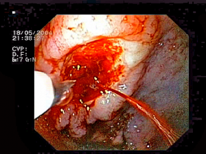
Image Credit: Jeremias, CC BY-SA 3.0, via Wikimedia Commons

Week 31 – Early TIPS in Cirrhosis with Variceal Bleeding

“Early Use of TIPS in Patients with Cirrhosis and Variceal Bleeding”

N Engl J Med. 2010 Jun 24;362(25):2370-9. [free full text]

Variceal bleeding is a major cause of morbidity and mortality in decompensated cirrhosis. The standard of care for an acute variceal bleed includes a combination of vasoactive drugs, prophylactic antibiotics, and endoscopic techniques (e.g. banding). Transjugular intrahepatic portosystemic shunt (TIPS) can be used to treat refractory bleeding. This 2010 trial sought to determine the utility of early TIPS during the initial bleed in high-risk patients when compared to standard therapy.

The trial enrolled cirrhotic patients (Child-Pugh class B or C with score ≤ 13) with acute esophageal variceal bleeding. All patients received endoscopic band ligation (EBL) or endoscopic injection sclerotherapy (EIS) at the time of diagnostic endoscopy. All patients also received vasoactive drugs (terlipressin, somatostatin, or octreotide). Patients were randomized to either TIPS performed within 72 hours after diagnostic endoscopy or to “standard therapy” by 1) treatment with vasoactive drugs with transition to nonselective beta blocker when patients were free of bleeding followed by 2) addition of isosorbide mononitrate to maximum tolerated dose, and 3) a second session of EBL at 7-14 days after the initial session (repeated q10-14 days until variceal eradication was achieved). The primary outcome was a composite of failure to control acute bleeding or failure to prevent “clinically significant” variceal bleeding (requiring hospital admission or transfusion) at 1 year after enrollment. Selected secondary outcomes included 1-year mortality, development of hepatic encephalopathy (HE), ICU days, and hospital LOS.

359 patients were screened for inclusion, but ultimately only 63 were randomized. Baseline characteristics were similar among the two groups except that the early TIPS group had a higher rate of patients with previous hepatic encephalopathy. The primary composite endpoint of failure to control acute bleeding or rebleeding within 1 year occurred in 14 of 31 (45%) patients in the pharmacotherapy-EBL group and in only 1 of 32 (3%) of the early TIPS group (p = 0.001). The 1-year actuarial probability of remaining free of the primary outcome was 97% in the early TIPS group vs. 50% in the pharmacotherapy-EBL group (ARR 47 percentage points, 95% CI 25-69 percentage points, NNT 2.1). Regarding mortality, at one year, 12 of 31 (39%) patients in the pharmacotherapy-EBL group had died, while only 4 of 32 (13%) in the early TIPS group had died (p = 0.001, NNT = 4.0). There were no group differences in prevalence of HE at one year (28% in the early TIPS group vs. 40% in the pharmacotherapy-EBL group, p = 0.13). Additionally, there were no group differences in 1-year actuarial probability of new or worsening ascites. There were also no differences in length of ICU stay or hospitalization duration.

Early TIPS in acute esophageal variceal bleeding, when compared to standard pharmacotherapy and endoscopic band ligation, improved control of index bleeding, reduced recurrent variceal bleeding at 1 year, and reduced all-cause mortality. Prior studies had demonstrated that TIPS reduced the rebleeding rate but increased the rate of hepatic encephalopathy without improving survival. As such, TIPS had only been recommended as a rescue therapy. Obviously, this study presents compelling data that challenge these paradigms. The authors note that in “patients with Child-Pugh class C or in class B with active variceal bleeding, failure to initially control the bleeding or early rebleeding contributes to further deterioration in liver function, which in turn worsens the prognosis and may preclude the use of rescue TIPS.” Authors at UpToDate note that, given the totality of evidence to date, the benefit of early TIPS in preventing rebleeding “is offset by its failure to consistently improve survival and increasing morbidity due to the development of liver failure and encephalopathy.” Today, TIPS remains primarily a salvage therapy for use in cases of recurrent bleeding despite standard pharmacotherapy and EBL. There may be a subset of patients in whom early TIPS is the ideal strategy, but further trials will be required to identify this subset.

Further Reading/References:
1. Wiki Journal Club []
2. 2 Minute Medicine []
3. UpToDate, “Prevention of recurrent variceal hemorrhage in patients with cirrhosis

Summary by Duncan F. Moore, MD

Week 20 – Omeprazole for Bleeding Peptic Ulcers

“Effect of Intravenous Omeprazole on Recurrent Bleeding After Endoscopic Treatment of Bleeding Peptic Ulcers”

N Engl J Med. 2000 Aug 3;343(5):310-6. [free full text]

Intravenous proton-pump inhibitor (PPI) therapy is a cornerstone of modern therapy for bleeding peptic ulcers. However, prior to this 2000 study by Lau et al., the role of PPIs in the prevention of recurrent bleeding after endoscopic treatment was unclear. At the time, re-bleeding rates after endoscopic treatment were noted to be approximately 15-20%. Although other studies had approached this question, no high-quality, large, blinded RCT had examined adjuvant PPI use immediately following endoscopic treatment.

The study enrolled patients who had a bleeding gastroduodenal ulcer visualized on endoscopy and in whom hemostasis was achieved following epinephrine injection and thermocoagulation. Enrollees were randomized to treatment with either omeprazole 80mg IV bolus followed by 8mg/hr infusion x72 hours then followed by omeprazole 20mg PO x8 weeks or to placebo bolus + drip x72 hours followed by omeprazole 20mg PO x8 weeks. The primary outcome was recurrent bleeding within 30 days. Secondary outcomes included recurrent bleeding within 72 hours, amount of blood transfused by day 30, hospitalization duration, and all-cause 30-day mortality.

120 patients were randomized to each arm. The trial was terminated early due to the finding on interim analysis of a significantly lower recurrent bleeding rate in the omeprazole arm. Bleeding re-occurred within 30 days in 8 (6.7%) omeprazole patients versus 27 (22.5%) placebo patients (HR 3.9, 95% CI 1.7-9.0; NNT 6.3). A Cox proportional-hazards model, when adjusted for size and location of ulcers, presence/absence of coexisting illness, and history of ulcer disease, revealed a similar hazard ratio (HR 3.9, 95% CI 1.7-9.1). Recurrent bleeding was most common during the first 72 hrs (4.2% of the omeprazole group versus 20% of the placebo group, RR 4.80, 95% CI 1.89-12.2, p<0.001). For a nice visualization of the early separation of re-bleeding rates, see the Kaplan-Meier curve in Figure 1. The mean number of units of blood transfused within 30 days was 2.7 ± 2.5 in the omeprazole group versus 3.5 ± 3.8 in the placebo group (p = 0.04). Regarding duration of hospitalization, 46.7% of omeprazole patients were admitted for < 5 days versus 31.7% of placebo patients (p = 0.02). Median stay was 4 days in the omeprazole group versus 5 days in the placebo group (p = 0.006). 4.2% of the omeprazole patients died within 30 days, whereas 10% of the placebo patients died (p = 0.13).

Treatment with intravenous omeprazole immediately following endoscopic intervention for bleeding peptic ulcer significantly reduced the rate of recurrent bleeding. This effect was most prominent within the first 3 days of therapy. This intervention also reduced blood transfusion requirements and shortened hospital stays. The presumed mechanism of action is increased gastric pH facilitating platelet aggregation. In 2018, the benefit of this intervention seems so obvious based on its description alone that one would not imagine that such a trial would be funded or published in such a high-profile journal. However, the annals of medicine are littered with now-discarded interventions that made sense from a theoretical or mechanistic perspective but were demonstrated to be ineffective or even harmful (e.g. pharmacologic suppression of ventricular arrhythmias post-MI or renal denervation for refractory HTN).

Today, bleeding peptic ulcers are treated with an IV PPI twice daily. Per UpToDate, meta-analyses have not shown a benefit of continuous PPI infusion over this IV BID dosing. However, per 2012 guidelines in the American Journal of Gastroenterology, patients with active bleeding or non-bleeding visible vessels should receive both endoscopic intervention and IV PPI bolus followed by infusion.

Further Reading/References:
1. Wiki Journal Club
2. 2 Minute Medicine
3. UpToDate, “Overview of the Treatment of Bleeding Peptic Ulcers”
4. Laine L, Jensen DM. “Management of patients with ulcer bleeding.” Am J Gastroenterol. 2012

Summary by Duncan F. Moore, MD

Image credit: Wesalius, CC BY 4.0, via Wikimedia Commons

Week 16 – MELD

“A Model to Predict Survival in Patients With End-Stage Liver Disease”

Hepatology. 2001 Feb;33(2):464-70. [free full text]

Prior to the adoption of the Model for End-Stage Liver Disease (MELD) score for the allocation of liver transplants, the determination of medical urgency was dependent on the Child-Pugh score. The Child-Pugh score was limited by the inclusion of two subjective variables (severity of ascites and severity of encephalopathy), limited discriminatory ability, and a ceiling effect of laboratory abnormalities. Stakeholders sought an objective, continuous, generalizable index that more accurately and reliably represented disease severity. The MELD score had originally been developed in 2000 to estimate the survival of patients undergoing TIPS. The authors of this 2001 study hypothesized that the MELD score would accurately estimate short-term survival in a wide range of severities and etiologies of liver dysfunction and thus serve as a suitable replacement measure for the Child-Pugh score in the determination of medical urgency in transplant allocation.

This study reported a series of four retrospective validation cohorts for the use of MELD in prediction of mortality in advanced liver disease. The index MELD score was calculated for each patient. Death during follow-up was assessed by chart review.

MELD score = 3.8*ln([bilirubin])+11.2*ln(INR)+9.6*ln([Cr])+6.4*(etiology: 0 if cholestatic or alcoholic, 1 otherwise)

The primary study outcome was the concordance c-statistic between MELD score and 3-month survival. The c-statistic is equivalent to the area under receiver operating characteristic (AUROC). Per the authors, “a c-statistic between 0.8 and 0.9 indicates excellent diagnostic accuracy and a c-statistic greater than 0.7 is generally considered as a useful test.” (See page 455 for further explanation.) There was no reliable comparison statistic (e.g. c-statistic of MELD vs. that of Child-Pugh in all groups).

C-statistic for 3-month survival in the four cohorts ranged from 0.78 to 0.87 (no 95% CIs exceeded 1.0). There was minimal improvement in the c-statistics for 3-month survival with the individual addition of spontaneous bacterial peritonitis, variceal bleed, ascites, and encephalopathy to the MELD score (see Table 4, highest increase in c-statistic was 0.03). When the etiology of liver disease was excluded from the MELD score, there was minimal change in the c-statistics (see Table 5, all paired CIs overlap). C-statistics for 1-week mortality ranged from 0.80 to 0.95.

In conclusion, the MELD score is an excellent predictor of short-term mortality in patients with end-stage liver disease of diverse etiology and severity. Despite the retrospective nature of this study, this study represented a significant improvement upon the Child-Pugh score in determining medical urgency in patients who require liver transplant. In 2002, the United Network for Organ Sharing (UNOS) adopted a modified version of the MELD score for the prioritization of deceased-donor liver transplants in cirrhosis. Concurrent with the 2001 publication of this study, Wiesner et al. performed a prospective validation of the use of MELD in the allocation of liver transplantation. When published in 2003, it demonstrated that MELD score accurately predicted 3-month mortality among patients with chronic liver disease on the waitlist. The MELD score has also been validated in other conditions such as alcoholic hepatitis, hepatorenal syndrome, and acute liver failure (see UpToDate). Subsequent additions to the MELD score have come out over the years. In 2006, the MELD Exception Guidelines offered extra points for severe comorbidities (e.g HCC, hepatopulmonary syndrome). In January 2016, the MELDNa score was adopted and is now used for liver transplant prioritization.

References and Further Reading:
1. “A model to predict poor survival in patients undergoing transjugular intrahepatic portosystemic shunts” (2000)
2. MDCalc “MELD Score”
3. Wiesner et al. “Model for end-stage liver disease (MELD) and allocation of donor livers” (2003)
4. Freeman Jr. et al. “MELD exception guidelines” (2006)
5. 2 Minute Medicine
6. UpToDate “Model for End-stage Liver Disease (MELD)”

Image Credit: Ed Uthman, CC-BY-2.0, via WikiMedia Commons

Week 2 – Albumin in SBP

“Effect of Intravenous Albumin on Renal Impairment and Mortality in Patients with Cirrhosis and Spontaneous Bacterial Peritonitis”

N Engl J Med. 1999 Aug 5;341(6):403-9. [free full text]

Renal failure commonly develops in the setting of SBP, and its development is a sensitive predictor of in-hospital mortality. The renal impairment is thought to stem from decreased effective arterial blood volume secondary to the systemic inflammatory response to the infection. In our current practice, there are certain circumstances in which we administer albumin early in the SBP disease course in order to reduce the risk of renal failure and mortality. Ultimately, our current protocol originated from the 1999 study of albumin in SBP by Sort et al.

The trial enrolled adults with SBP and randomized them to treatment with either cefotaxime and albumin infusion 1.5 gm/kg within 6hrs of enrollment, followed by 1 gm/kg on day 3 or cefotaxime alone. The primary outcome was the development of “renal impairment” (a “nonreversible” increase in BUN or Cr by more than 50% to a value greater than 30 mg/dL or 1.5 mg/dL, respectively) during hospitalization. The secondary outcome was in-hospital mortality.

126 patients were randomized. Both groups had similar baseline characteristics, and both had similar rates of resolution of infection. Renal impairment occurred in 10% of the albumin group and 33% of the cefotaxime-alone group (p=0.02). In-hospital mortality was 10% in the albumin group and 29% in the cefotaxime-alone group (p=0.01). 78% of patients that developed renal impairment died in-hospital, while only 3% of patients who did not develop renal impairment died. Plasma renin activity was significantly higher on days 3, 6, and 9 in the cefotaxime-alone group than in the albumin group, while there were no significant differences in MAP among the two groups at those time intervals. Multivariate analysis of all trial participants revealed that baseline serum bilirubin and creatinine were independent predictors of the development of renal impairment.

In conclusion, albumin administration reduces renal impairment and improves mortality in patients with SBP. The findings of this landmark trial were refined by a brief 2007 report by Sigal et al. entitled “Restricted use of albumin for spontaneous bacterial peritonitis.” “High-risk” patients, identified by baseline serum bilirubin of ≥ 4.0 mg/dL or Cr ≥ 1.0 mg/dL were given the intervention of albumin 1.5gm/kg on day 1 and 1gm/kg on day 3, and low-risk patients were not given albumin. None of the 15 low-risk patients developed renal impairment or died, whereas 12 of 21 (57%) of the high-risk group developed renal impairment, and 5 of the 21 (24%) died. The authors conclude that patients with bilirubin < 4.0 and Cr < 1.0 did not need scheduled albumin in the treatment of SBP. The current (2012) American Association for the Study of Liver Diseases guidelines for the management of adult patients with ascites due to cirrhosis do not definitively recommend criteria for albumin administration in SBP. Instead they summarize the aforementioned two studies. A 2013 meta-analysis of four reports/trials (including the two above) concluded that albumin infusion reduced renal impairment and improved mortality with pooled odds ratios approximately commensurate with those of the 1999 study by Sort et al. Ultimately, the current recommended practice per expert opinion is to perform albumin administration per the protocol outlined by Sigal et al. (2007).

References / Further Reading:
1. AASLD Guidelines for Management of Adult Patients with Ascites Due to Cirrhosis (skip to page 77)
2. Sigal et al. “Restricted use of albumin for spontaneous bacterial peritonitis.” Gut 2007.
3. Meta-analysis: “Albumin infusion improves outcomes of patients with spontaneous bacterial peritonitis: a meta-analysis of randomized trials”
4. Wiki Journal Club
5. 2 Minute Medicine

Summary by Duncan F. Moore, MD

Week 49 – STOPAH

“Prednisolone or Pentoxifylline for Alcohol Hepatitis”

aka the Steroids or Pentoxifylline for Alcoholic Hepatitis (STOPAH) trial

N Engl J Med. 2015 Apr 23;372(17):1619-28. [free full text]

Severe alcoholic hepatitis is associated with short-term mortality as high as 30%. Treatment of alcoholic hepatitis with corticosteroids has been extensively studied and debated extensively. Prior to this 2010 study, an analysis of the five largest studies of glucocorticoid treatment in alcoholic hepatitis concluded that there was a significant mortality benefit at 28 days among patients with severe disease. Similarly, the nonselective phosphodiesterase inhibitor pentoxifylline has been evaluated in alcoholic hepatitis. One of four RCTs showed a significant benefit, but two meta-analyses have not concluded that there is any benefit. The authors of the 2010 STOPAH trial sought to evaluate both therapies compared to placebos in a 2-by-2 factorial design.

Population: adults with a clinical diagnosis of alcoholic hepatitis, average alcohol consumption > 80 gm/day in men or 60 gm/day in women, total bilirubin > 4.7mg/dL, and a Maddrey discriminant function ≥ 32

Intervention / Comparison: patients were randomized to one of the following four groups for 28 days of treatment

  • prednisolone-matched placebo daily + pentoxifylline-matched placebo TID
  • prednisolone 40mg daily + pentoxifylline-matched placebo TID
  • prednisolone-matched placebo daily + pentoxifylline 400mg TID
  • prednisolone 40mg daily + pentoxifylline 400mg TID

Outcome:
Primary – 28-day mortality
Secondary – mortality or liver transplant at 90 days and at 1 year

Results:
Regarding randomization of the 1103 patients, 276 were randomized to placebo-placebo, 277 to prednisolone-placebo, 276 to pentoxifylline-placebo, and 274 to prednisolone-pentoxifylline. The trial was stopped early due to “limitations on funding.” However, all enrolled patients completed at least 28 days of follow-up. 33 patients were unable to complete 90-day and 1-year follow up due to termination of the trial.

At 28 days, 45 of 269 (17%) of placebo-placebo patients, 38 of 266 (14%) of prednisolone-placebo patients, 50 of 258 (19%) of pentoxifylline-placebo patients, and 35 of 260 (13%) of prednisolone-pentoxifylline patients had died. The odds ratio for 28-day mortality among patients treated with prednisolone was 0.72 (95% CI 0.52-1.01, p = 0.06), and the odds ratio for patients treated with pentoxifylline was 1.07 (95% CI 0.77-1.49, p = 0.69).

Similarly, neither treatment was found to influence 90-day or 1-year mortality or liver transplantation (see Table 2).

Infection occurred in 13% of patients who received prednisolone versus 7% of patients who did not receive prednisolone.

Implication/Discussion:
In patients with severe alcoholic hepatitis, neither prednisolone nor pentoxifylline reduced morality risk at 28 days. Additionally, neither drug reduced the combined secondary endpoint of mortality or liver transplantation at 90 days or 1 year.

This was a well-designed, randomized, double-blind, double-placebo-controlled trial.

A notable limitation was this trial’s reliance on the clinical diagnosis of alcohol hepatitis, rather than tissue diagnosis. This may have reduced the power of the trial with respect to detecting a treatment effect. Contemporary authors also noted that harm may have come to study patients due to a lack of tapering of prednisolone at the end of the 28 days of treatment.

A 2015 meta-analysis that included the STOPAH trial concluded that prednisolone treatment reduced 28-day mortality.

Despite the negative results of this specific trial, corticosteroid treatment has remained a mainstay of the treatment of severe alcoholic hepatitis.

The generally accepted practice, as summarized by UpToDate, is treatment with prednisolone 40mg PO daily for 28 days in patients with discriminant function ≥ 32. (Prednisolone is preferred over prednisone, because prednisone requires conversion in the liver to its active form prednisolone, and such conversion can be impaired in liver dysfunction.) Therapy should be terminated early after 7 days if patients fail to show improvement (either by parameters such as bilirubin or discriminant function, or by improvement in the Lille score).

Further Reading/References:
1. Wiki Journal Club
2. 2 Minute Medicine
3. UpToDate, “Management and prognosis of alcoholic hepatitis”
4. American College of Gastroenterology, “ACG Clinical Guideline: Alcoholic Liver Disease” (2018)
5. European Association for Study of the Liver (EASL), “EASL Clinical Practice Guidelines: Management of Alcoholic Liver Disease” (2012)

Summary by Duncan F. Moore, MD

Week 46 – Transfusion Strategies for Upper GI Bleeding

“Transfusion Strategies for Acute Upper Gastrointestinal Bleeding”

N Engl J Med. 2013 Jan 3;368(1):11-21. [free full text]

A restrictive transfusion strategy of 7 gm/dL was established following the previously discussed 1999 TRICC trial. Notably, both TRICC and its derivative study TRISS excluded patients who had an active bleed. In 2013, Villanueva et al. performed a study to establish whether there was benefit to a restrictive transfusion strategy in patients with acute upper GI bleeding.

Population: adults with hematemesis (or bloody nasogastric aspirate), melena, or both; selected consecutively at a single-center in Spain

Notable exclusion criteria: a clinical Rockall score* of 0 with a hemoglobin level higher than 12g/dL, massive exsanguinating bleeding, lower GIB, patient refusal of blood transfusion, ACS, stroke/TIA, transfusion within 90 days, recent trauma or surgery

*The Rockall score is a system to assess risk for further bleeding or death on a scale from 0-11. Higher scores (3-11) indicate higher risk. Of the 648 patients excluded, the most common reason for exclusion (n = 329) was low risk of bleeding.

Intervention: restrictive transfusion strategy (transfusion threshold Hgb = 7.0 gm/dL) [n = 444]

Comparison: liberal transfusion strategy (transfusion threshold Hgb = 9.0 gm/dL) [n = 445]

During randomization, patients were stratified by presence or absence of cirrhosis.

As part of the study design, all patients underwent emergent EGD within 6 hours and received relevant hemostatic intervention depending on the cause of bleeding.

Outcome:
Primary outcome: 45-day mortality

Secondary outcomes, selected:

  • Incidence of further bleeding associated with hemodynamic instability or hemoglobin drop > 2 gm/dL in 6 hours
  • Incidence and number of RBC transfusions
  • Other products and fluids transfused
  • Hgb level at nadir, discharge, and 45 days

Subgroup analyses: Patients were stratified by presence of cirrhosis and corresponding Child-Pugh class, variceal bleeding, and peptic ulcer bleeding. An additional subgroup analysis was performed to evaluate changes in hepatic venous pressure gradient between the two strategies.


Results
:
The primary outcome of 45-day mortality was lower in the restrictive strategy (5% vs. 9%; HR 0.55, 95% CI 0.33-0.92; p = 0.02; NNT = 24.8). In subgroup analysis, this finding remained consistent for patients who had Child-Pugh class A or B but was not statistically significant among patients who had Class C. Further stratification for variceal bleeding and peptic ulcer disease did not make a difference in mortality.

Secondary outcomes:
Rates of further bleeding events and RBC transfusion, as well as number of products transfused, were lower in the restrictive strategy. Subgroup analysis demonstrated that rates of re-bleeding were lower in Child-Pugh class A and B but not in C. As expected, the restrictive strategy also resulted in the lowest hemoglobin levels at 24 hours. Hemoglobin levels among patients in the restrictive strategy were lower at discharge but were not significantly different from the liberal strategy at 45 days. There was no group difference in amount of non-RBC blood products or colloid/crystalloid transfused. Patients in the restrictive strategy experienced fewer adverse events, particularly transfusion reactions such as transfusion-associated circulatory overload and cardiac complications. Patients in the liberal-transfusion group had significant post-transfusion increases in mean hepatic venous pressure gradient following transfusion. Such increases were not seen in the restrictive-strategy patients.


Implication/Discussion
:
In patients with acute upper GI bleeds, a restrictive strategy with a transfusion threshold 7 gm/dL reduces 45-day mortality, the rate and frequency of transfusions, and the rate of adverse reactions, relative to a liberal strategy with a transfusion threshold of 9 gm/dL.

In their discussion, the authors hypothesize that the “harmful effects of transfusion may be related to an impairment of hemostasis. Transfusion may counteract the splanchnic vascoconstrictive response caused by hypovolemia, inducing an increase in splanchnic blood flow and pressure that may impair the formation of clots. Transfusion may also induce abnormalities in coagulation properties.”

Subgroup analysis suggests that the benefit of the restrictive strategy is less pronounced in patients with more severe hepatic dysfunction. These findings align with prior studies in transfusion thresholds for critically ill patients. However, the authors note that the results conflict with studies in other clinical circumstances, specifically in the pediatric ICU and in hip surgery for high-risk patients.

There are several limitations to this study. First, its exclusion criteria limit its generalizability. Excluding patients with massive exsanguination is understandable given lack of clinical equipoise; however, this choice allows too much discretion with respect to the definition of a massive bleed. (Note that those excluded due to exsanguination comprised only 39 of 648.) Lack of blinding was a second limitation. Potential bias was mitigated by well-defined transfusion protocols. Additionally, there a higher incidence of transfusion-protocol violations in the restrictive group, which probably biased results toward the null. Overall, deviations from the protocol occurred in fewer than 10% of cases.


Further Reading/References
:
1. Transfusion Strategies for Acute Upper GI Bleeding @ Wiki Journal Club
2. 2 Minute Medicine
3. TRISS @ Wiki Journal Club

Summary by Gordon Pelegrin, MD

Week 44 – Early TIPS in Cirrhosis with Variceal Bleeding

“Early Use of TIPS in Patients with Cirrhosis and Variceal Bleeding”

N Engl J Med. 2010 Jun 24;362(25):2370-9. [free full text]

Variceal bleeding is a major cause of morbidity and mortality in decompensated cirrhosis. The standard of care for an acute variceal bleed includes a combination of vasoactive drugs, prophylactic antibiotics, and endoscopic techniques (e.g. banding). Transjugular intrahepatic portosystemic shunt (TIPS) can be used to treat refractory bleeding. This 2010 trial sought to determine the utility of early TIPS during the initial bleed in high-risk patients, when compared to standard therapy.

Population: cirrhotic patients with acute esophageal variceal bleeding, either Child-Pugh class C with score 10-13 or class B (score 7-9) with active bleeding at diagnostic endoscopy

Notable exclusion criteria: Child-Pugh score > 13, age > 75, HCC that did not meet transplantation criteria, bleeding gastric varices, total portal vein thrombosis, prior TIPS

All patients received endoscopic band ligation (EBL) or endoscopic injection sclerotherapy (EIS) at the time of diagnostic endoscopy. All patients also received vasoactive drugs (terlipressin, somatostatin, or octreotide).

Intervention: TIPS performed within 72 hours after diagnostic endoscopy

Comparison: 1) treatment with vasoactive drugs with transition to nonselective beta blocker when patients free of bleeding followed by 2) addition of isosorbide mononitrate to maximum tolerated dose, and 3) a second session of EBL at 7-14 days after the initial session (repeated q10-14 days until variceal eradication was achieved)

Outcome:
Primary – composite of failure to control acute bleeding or failure to prevent “clinically significant” variceal bleeding (requiring hospital admission or transfusion) at 1 year after enrollment

Secondary, selected

  • mortality at 1 year
  • failure to control acute bleeding
  • early rebleeding (at 5 days and 6 weeks)
  • rate of development of hepatic encephalopathy (HE)
  • ICU days, time in hospital


Results:
359 patients were screened for inclusion, but ultimately only 63 were randomized. Baseline characteristics were similar among the two groups except that the early TIPS group had a higher rate of patients with previous hepatic encephalopathy. Among early TIPS patients, the mean portal pressure dropped from 20.2±7 mmHg to 6.2±3 mmHg.

The primary composite endpoint of failure to control acute bleeding or rebleeding within 1 year occurred in 14 of 31 (45%) patients in the pharmacotherapy-EBL group and in only 1 of 32 (3%) of the early TIPS group (p = 0.001). The 1-year actuarial probability of remaining free of the primary outcome was 97% in the early TIPS group vs. 50% in the pharmacotherapy-EBL group (ARR 47 percentage points, 95% CI 25-69 percentage points, NNT 2.1).

Regarding mortality, at one year, 12 of 31 (39%) patients in the pharmacotherapy-EBL group had died, while only 4 of 32 (13%) in the early TIPS group had died (p = 0.001, NNT = 4.0).

Regarding HE: the 1-year actuarial probability of HE was 28% in the early TIPS group vs. 40% in the pharmacotherapy-EBL group (p = 0.13). Most of the episodes of HE occurred during the index bleed. Following discharge from index hospitalization, the 1-year risk of additional HE episodes was 10% in the pharmacotherapy-EBL group and 19% in the early TIPS group (p = 0.80).

There were no group differences in 1-year actuarial probability of new or worsening ascites.

There were no group differences in length of ICU stay or hospitalization duration.

Implication/Discussion:
Early TIPS in acute esophageal variceal bleeding, when compared to standard pharmacotherapy and endoscopic band ligation, improved control of index bleeding, reduced recurrent variceal bleeding at 1 year, and reduced all-cause mortality.

Prior studies had demonstrated that TIPS reduced the rebleeding rate but increased the rate of hepatic encephalopathy without improving survival. As such, TIPS had only been recommended as a rescue therapy. Obviously, this study presents compelling data that challenges these paradigms.

The authors note that in “patients with Child-Pugh class C or in class B with active variceal bleeding, failure to initially control the bleeding or early rebleeding contributes to further deterioration in liver function, which in turn worsens the prognosis and may preclude the use of rescue TIPS.”

Authors at UpToDate note that, given the totality of evidence to date, the benefit of early TIPS in preventing rebleeding “is offset by its failure to consistently improve survival and increasing morbidity due to the development of liver failure and encephalopathy.” Today, TIPS remains primarily a salvage therapy for use in cases of recurrent bleeding despite standard pharmacotherapy and EBL. There may be a subset of patients in whom early TIPS is the ideal strategy, but further trials will be required to identify this subset.


Further Reading/References
:
1. Wiki Journal Club
2. 2 Minute Medicine
3. UpToDate, “Prevention of recurrent variceal hemorrhage in patients with cirrhosis”

Summary by Duncan F. Moore, MD

Week 32 – Rifaximin Treatment in Hepatic Encephalopathy

“Rifaximin Treatment in Hepatic Encephalopathy”

N Engl J Med. 2010 Mar25;362(12):1071-81. [free full text]

As we are well aware at Georgetown, hepatic encephalopathy (HE) is highly prevalent among patients with cirrhosis, and admissions for recurrent HE place a significant burden on the medical system. The authors of this study note that HE is thought to result from “the systemic accumulation of gut-derived neurotoxins, especially ammonia, in patients with impaired liver function and portosystemic shunting.” Lactulose is considered the standard of care for the prevention of HE. It is thought to decrease the absorption of ammonia in the gut lumen through its cathartic effects and by alteration of colonic pH. The minimally absorbable oral antibiotic rifaximin is thought to further reduce ammonia production through direct antibacterial effects within the gut lumen. Thus the authors of this pivotal 2010 study sought to determine the additive effect of daily rifaximin prophylaxis in the prevention of HE.

Population: adults with cirrhosis and 2+ episodes of overt HE during the past 6 months

Notable exclusion criteria: West Haven Criteria score of 2+ at enrollment, MELD score > 25, baseline Cr > 2.0, or if prior qualifying HE episodes were 2/2 GIB, medication effect, or renal failure

Intervention: rifaximin 550mg PO BID x6 months

Comparison: placebo 550mg PO BID x6 months

Outcomes:

Primary: time to first breakthrough episode of HE (West Haven Score of 2+ or West Haven Score 0 –> 1 with worsening asterixis)
Secondary

  • time to first hospitalization involving HE
  • adverse events, including those “possibly related to infection”

Results:
299 patients were randomized. 140 and 159 patients were assigned to rifaximin and placebo, respectively. Baseline characteristics were similar among the two groups. Lactulose use prior to and during the study was similar in both groups at approximately 91%.

Breakthrough HE occurred in 31 (22.1%) of the rifaximin patients and 73 (45.9%) of the placebo patients [HR 0.42, 95% CI 0.28-0.64, p<0.001, absolute risk reduction 23.7%, NNT = 4.2]. This result was consistent within all tested subgroups, except patients with MELD score 19-24 and patients who were not using lactulose at baseline (see Figure 3).

Hospitalization involving HE occurred in 19 (13.6%) of the rifaximin patients and 36 (22.6%) of the placebo patients [HR 0.50, 95% CI 0.29-0.87, p = 0.01, absolute risk reduction 9.1%, NNT = 11.0].

There were no differences in adverse events among the two treatment groups.

Implication/Discussion:
Prophylactic rifaximin reduced the incidence of recurrent hepatic encephalopathy and its resultant hospitalizations.

This landmark trial showed a clear treatment benefit with implied savings in healthcare utilization costs associated with HE recurrences and hospitalizations. This marked effect was demonstrated even in the setting of relatively good (91%) lactulose adherence in both treatment arms prior to and throughout the trial.

On the day this trial was published in 2010, the FDA approved rifaximin for “reduction in risk of overt hepatic encephalopathy recurrence” in adults.

Because rifaximin is not generic and remains quite expensive, its financial utility is limited from an insurance company’s perspective. There is no other comparable nonabsorbable antibiotic for this indication.

UpToDate suggests starting with lactulose therapy and then adding a nonabsorbable antibiotic, such as rifaximin, both for the treatment of overt hepatic encephalopathy and for the prevention of recurrent hepatic encephalopathy. In practice, most insurance companies will require a prior authorization for outpatient rifaximin treatment, but in my recent experience this process has been perfunctory and easy.

Further Reading/References:
1. ClinicalTrials.gov, NCT00298038
2. FDA, NDA approval letter for Xifaxan (rifaximin)

Summary by Duncan F. Moore, MD

Week 30 – Omeprazole for Bleeding Peptic Ulcers

“Effect of Intravenous Omeprazole on Recurrent Bleeding After Endoscopic Treatment of Bleeding Peptic Ulcers”

N Engl J Med. 2000 Aug 3;343(5):310-6. [free full text]

Intravenous proton-pump inhibitor (PPI) therapy is a cornerstone of modern therapy for bleeding peptic ulcers. However, prior to this 2000 study by Lau et al., the role of PPIs in the prevention of recurrent bleeding after endoscopic treatment was unclear. At the time, re-bleeding rates after endoscopic treatment were noted to be approximately 15-20%. Although other studies had approached this question, no high-quality, large, blinded RCT had examined adjuvant PPI use immediately following endoscopic treatment.

Population: patients with bleeding gastroduodenal ulcer visualized on endoscopy in whom hemostasis was achieved following epinephrine injection and thermocoagulation (consecutive patients, single center in Hong Kong)

Intervention: omeprazole 80mg IV bolus followed by 8mg/hr infusion x72 hrs, followed by omeprazole 20mg PO x8 wks

Comparison: placebo bolus + drip x72 hrs, followed by omeprazole 20mg PO x8 wks

Outcome:
Primary – Recurrent bleeding within 30 days

Secondary

  1. Recurrent bleeding within 72 hrs
  2. Mean number of units of blood transfused within 30 days
  3. Duration of hospitalization
  4. All-cause mortality at 30 days


Results
:
120 patients were randomized to each arm. The two groups had similar baseline characteristics, including ulcer-specific characteristics. The trial was terminated early due to the finding on interim analysis of a significantly lower recurrent bleeding rate in the omeprazole arm.

Bleeding re-occurred within 30 days in 8 (6.7%) omeprazole patients versus 27 (22.5%) placebo patients (HR 3.9, 95% CI 1.7-9.0; NNT 6.3). A Cox proportional-hazards model, when adjusted for size and location of ulcers, presence/absence of coexisting illness, and history of ulcer disease, revealed a similar hazard ratio (HR 3.9, 95% CI 1.7-9.1).

Recurrent bleeding was most common during the first 72 hrs (4.2% of the omeprazole group versus 20% of the placebo group, RR 4.80, 95% CI 1.89-12.2, p<0.001). For a nice visualization of the early separation of re-bleeding rates, see the Kaplan-Meier curve in Figure 1.

The mean number of units of blood transfused within 30 days was 2.7 ± 2.5 in the omeprazole group versus 3.5 ± 3.8 in the placebo group (p = 0.04). The number of units transfused after the initial endoscopic treatment was 1.7 ± 1.9 in the omeprazole group versus 2.4 ± 3.2 in the placebo group (p = 0.03).

Regarding duration of hospitalization, 46.7% of omeprazole patients were admitted for < 5 days versus 31.7% of placebo patients (p = 0.02). Median stay was 4 days in the omeprazole group versus 5 days in the placebo group (p = 0.006).

4.2% of the omeprazole patients died within 30 days, whereas 10% of the placebo patients died (p = 0.13).

Implication/Discussion:
Treatment with intravenous omeprazole immediately following endoscopic intervention for bleeding peptic ulcer significantly reduced the rate of recurrent bleeding. This effect was most prominent within the first 3 days of therapy. This intervention also reduced blood transfusion requirements and shortened hospital stays.

The presumed mechanism of action is increased gastric pH facilitating platelet aggregation.

In 2018, the benefit of this intervention seems so obvious based on its description alone, that one would imagine that such a trial would not be funded or published in such a high-profile journal. However, the annals of medicine are littered with now-discarded interventions that made sense from a theoretical or mechanistic perspective but were demonstrated to be ineffective or even harmful (e.g. pharmacologic suppression of ventricular arrhythmias post-MI or renal denervation for refractory HTN).

Today, bleeding peptic ulcers are treated with an IV PPI twice daily. Per UpToDate, meta-analyses have not shown a benefit of continuous PPI infusion over this IV BID dosing. However, per 2012 guidelines in the American Journal of Gastroenterology, patients with active bleeding or non-bleeding visible vessels should receive both endoscopic intervention and IV PPI bolus followed by infusion.


Further Reading/References
:
1. Wiki Journal Club
2. 2 Minute Medicine
3. UpToDate, “Overview of the Treatment of Bleeding Peptic Ulcers”
4. Laine L, Jensen DM. “Management of patients with ulcer bleeding.” Am J Gastroenterol. 2012.

Summary by Duncan F. Moore, MD