Week 30 – Bicarbonate and Progression of CKD

“Bicarbonate Supplementation Slows Progression of CKD and Improves Nutritional Status”

J Am Soc Nephrol. 2009 Sep;20(9):2075-84. [free full text]

Metabolic acidosis is a common complication of advanced CKD. Some animal models of CKD have suggested that worsening metabolic acidosis is associated with worsening proteinuria, tubulointerstitial fibrosis, and acceleration of decline of renal function. Short-term human studies have demonstrated that bicarbonate administration reduces protein catabolism and that metabolic acidosis is an independent risk factor for acceleration of decline of renal function. However, until this 2009 study by de Brito-Ashurst et al., there were no long-term studies demonstrating the beneficial effects of oral bicarbonate administration on CKD progression and nutritional status.

The study enrolled CKD patients with CrCl 15-30ml/min and plasma bicarbonate 16-20 mEq/L and randomized them to treatment with either sodium bicarbonate 600mg PO TID (with protocolized uptitration to achieve plasma HCO3  ≥ 23 mEq/L) for 2 years, or to routine care. The primary outcomes were: 1) the decline in CrCl at 2 years, 2) “rapid progression of renal failure” (defined as decline of CrCl > 3 ml/min per year), and 3) development of ESRD requiring dialysis. Secondary outcomes included 1) change in dietary protein intake, 2) change in normalized protein nitrogen appearance (nPNA), 3) change in serum albumin, and 4) change in mid-arm muscle circumference.

134 patients were randomized, and baseline characteristics were similar among the two groups. Serum bicarbonate levels increased significantly in the treatment arm. (See Figure 2.) At two years, CrCl decline was 1.88 ml/min in the treatment group vs. 5.93 ml/min in the control group (p < 0.01). Rapid progression of renal failure was noted in 9% of intervention group vs. 45% of the control group (RR 0.15, 95% CI 0.06–0.40, p < 0.0001, NNT = 2.8), and ESRD developed in 6.5% of the intervention group vs. 33% of the control group (RR 0.13, 95% CI 0.04–0.40, p < 0.001; NNT = 3.8). Regarding nutritional status, dietary protein intake increased in the treatment group relative to the control group (p < 0.007). Normalized protein nitrogen appearance decreased in the treatment group and increased in the control group (p < 0.002). Serum albumin increased in the treatment group but was unchanged in the control group, and mean mid-arm muscle circumference increased by 1.5 cm in the intervention group vs. no change in the control group (p < 0.03).

In conclusion, oral bicarbonate supplementation in CKD patients with metabolic acidosis reduces the rate of CrCl decline and progression to ESRD and improves nutritional status. Primarily on the basis of this study, the KDIGO 2012 guidelines for the management of CKD recommend oral bicarbonate supplementation to maintain serum bicarbonate within the normal range (23-29 mEq/L). This is a remarkably cheap and effective intervention. Importantly, the rates of adverse events, particularly worsening hypertension and increasing edema, were unchanged among the two groups. Of note, sodium bicarbonate induces much less volume expansion than a comparable sodium load of sodium chloride.

In their discussion, the authors suggest that their results support the hypothesis of Nath et al. (1985) that “compensatory changes [in the setting of metabolic acidosis] such as increased ammonia production and the resultant complement cascade activation in remnant tubules in the declining renal mass [are] injurious to the tubulointerstitium.” The hypercatabolic state of advanced CKD appears to be mitigated by bicarbonate supplementation. The authors note that “an optimum nutritional status has positive implications on the clinical outcomes of dialysis patients, whereas [protein-energy wasting] is associated with increased morbidity and mortality.”

Limitations to this trial include its open-label, no-placebo design. Also, the applicable population is limited by study exclusion criteria of morbid obesity, overt CHF, and uncontrolled HTN.

Further Reading:
1. Nath et al. “Pathophysiology of chronic tubulo-interstitial disease in rats: Interactions of dietary acid load, ammonia, and complement component-C3” (1985)
2. KDIGO 2012 Clinical Practice Guideline for the Evaluation and Management of Chronic Kidney Disease (see page 89)
3. UpToDate, “Pathogenesis, consequences, and treatment of metabolic acidosis in chronic kidney disease”
Week 25 – ALLHAT

“Major Outcomes in High-Risk Hypertensive Patients Randomized to Angiotensin-Converting Enzyme Inhibitor or Calcium Channel Blocker vs. Diuretic”

The Antihypertensive and Lipid-Lowering Treatment to Prevent Heart Attack Trial (ALLHAT)

JAMA. 2002 Dec 18;288(23):2981-97. [free full text]

Hypertension is a ubiquitous disease, and the cardiovascular and mortality benefits of BP control have been well described. However, as the number of available antihypertensive classes proliferated in the past several decades, a head-to-head comparison of different antihypertensive regimens was necessary to determine the optimal first-step therapy. The 2002 ALLHAT trial was a landmark trial in this effort.

Population:
33,357 patients aged 55 years or older with hypertension and at least one other coronary heart disease (CHD) risk factor (previous MI or stroke, LVH by ECG or echo, T2DM, current cigarette smoking, HDL < 35 mg/dL, or documentation of other atherosclerotic cardiovascular disease (CVD)). Notable exclusion criteria: history of hospitalization for CHF, history of treated symptomatic CHF, or known LVEF < 35%.

Intervention:
Prior antihypertensives were discontinued upon initiation of the study drug. Patients were randomized to one of three study drugs in a double-blind fashion. Study drugs and additional drugs were added in a step-wise fashion to achieve a goal BP < 140/90 mmHg.

Step 1: titrate assigned study drug

  • chlorthalidone: 12.5 –> 5 (sham titration) –> 25 mg/day
  • amlodipine: 2.5 –> 5 –>  10 mg/day
  • lisinopril: 10 –> 20 –> 40 mg/day

Step 2: add open-label agents at treating physician’s discretion (atenolol, clonidine, or reserpine)

  • atenolol: 25 to 100 mg/day
  • reserpine: 0.05 to 0.2 mg/day
  • clonidine: 0.1 to 0.3 mg BID

Step 3: add hydralazine 25 to 100 mg BID

Comparison:
Pairwise comparisons with respect to outcomes of chlorthalidone vs. either amlodipine or lisinopril. A doxazosin arm existed initially, but it was terminated early due to an excess of CV events, primarily driven by CHF.

Outcomes:
Primary –  combined fatal CAD or nonfatal MI

Secondary

  • all-cause mortality
  • fatal and nonfatal stroke
  • combined CHD (primary outcome, PCI, or hospitalized angina)
  • combined CVD (CHD, stroke, non-hospitalized treated angina, CHF [fatal, hospitalized, or treated non-hospitalized], and PAD)

Results:
Over a mean follow-up period of 4.9 years, there was no difference between the groups in either the primary outcome or all-cause mortality.

When compared with chlorthalidone at 5 years, the amlodipine and lisinopril groups had significantly higher systolic blood pressures (by 0.8 mmHg and 2 mmHg, respectively). The amlodipine group had a lower diastolic blood pressure when compared to the chlorthalidone group (0.8 mmHg).

When comparing amlodipine to chlorthalidone for the pre-specified secondary outcomes, amlodipine was associated with an increased risk of heart failure (RR 1.38; 95% CI 1.25-1.52).

When comparing lisinopril to chlorthalidone for the pre-specified secondary outcomes, lisinopril was associated with an increased risk of stroke (RR 1.15; 95% CI 1.02-1.30), combined CVD (RR 1.10; 95% CI 1.05-1.16), and heart failure (RR 1.20; 95% CI 1.09-1.34). The increased risk of stroke was mostly driven by 3 subgroups: women (RR 1.22; 95% CI 1.01-1.46), blacks (RR 1.40; 95% CI 1.17-1.68), and non-diabetics (RR 1.23; 95% CI 1.05-1.44). The increased risk of CVD was statistically significant in all subgroups except in patients aged less than 65. The increased risk of heart failure was statistically significant in all subgroups.

Discussion:
In patients with hypertension and one risk factor for CAD, chlorthalidone, lisinopril, and amlodipine performed similarly in reducing the risks of fatal CAD and nonfatal MI.

The study has several strengths: a large and diverse study population, a randomized, double-blind structure, and the rigorous evaluation of three of the most commonly prescribed “newer” classes of antihypertensives. Unfortunately, neither an ARB nor an aldosterone antagonist was included in the study. Additionally, the step-up therapies were not reflective of contemporary practice. (Instead, patients would likely be prescribed one or more of the primary study drugs.)

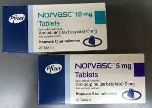
The ALLHAT study is one of the hallmark studies of hypertension and has played an important role in hypertension guidelines since it was published. Following the publication of ALLHAT, thiazide diuretics became widely used as first line drugs in the treatment of hypertension. The low cost of thiazides and their limited side-effect profile are particularly attractive class features. While ALLHAT looked specifically at chlorthalidone, in practice the positive findings were attributed to HCTZ, which has been more often prescribed. The authors of ALLHAT argued that the superiority of thiazides was likely a class effect, but according to the analysis at Wiki Journal Club, “there is little direct evidence that HCTZ specifically reduces the incidence of CVD among hypertensive individuals.” Furthermore, a 2006 study noted that that HCTZ has worse 24-hour BP control than chlorthalidone due to a shorter half-life. The ALLHAT authors note that “since a large proportion of participants required more than 1 drug to control their BP, it is reasonable to infer that a diuretic be included in all multi-drug regimens, if possible.” The 2017 ACC/AHA High Blood Pressure Guidelines state that, of the four thiazide diuretics on the market, chlorthalidone is preferred because of a prolonged half-life and trial-proven reduction of CVD (via the ALLHAT study).

Further Reading / References:
1. 2017 ACC Hypertension Guidelines
2. Wiki Journal Club
3. 2 Minute Medicine
4. Ernst et al, “Comparative antihypertensive effects of hydrochlorothiazide and chlorthalidone on ambulatory and office blood pressure.” (2006)
5. Gillis Pharmaceuticals [https://www.youtube.com/watch?v=HOxuAtehumc]
6. Concepts in Hypertension, Volume 2 Issue 6

Summary by Ryan Commins MD

Image Credit: Kimivanil, CC BY-SA 4.0, via Wikimedia Commons

Week 18 – Early Palliative Care in NSCLC

“Early Palliative Care for Patients with Metastatic Non-Small-Cell Lung Cancer”

N Engl J Med. 2010 Aug 19;363(8):733-42 [free full text]

Ideally, palliative care improves a patient’s quality of life while facilitating appropriate usage of healthcare resources. However, initiating palliative care late in a disease course or in the inpatient setting may limit these beneficial effects. This 2010 study by Temel et al. sought to demonstrate benefits of early integrated palliative care on patient-reported quality-of-life (QoL) outcomes and resource utilization.

The study enrolled outpatients with metastatic NSCLC diagnosed < 8 weeks ago and ECOG performance status 0-2 and randomized them to either “early palliative care” (met with palliative MD/ARNP within 3 weeks of enrollment and at least monthly afterward) or to standard oncologic care. The primary outcome was the change in Trial Outcome Index (TOI) from baseline to 12 weeks.

TOI = sum of the lung cancer, physical well-being, and functional well-being subscales of the Functional Assessment of Cancer Therapy­–Lung (FACT-L) scale (scale range 0-84, higher score = better function)

Secondary outcomes included:

  1. change in FACT-L score at 12 weeks (scale range 0-136)
  2. change in lung cancer subscale of FACT-L at 12 weeks (scale range 0-28)
  3. “aggressive care,” meaning one of the following: chemo within 14 days before death, lack of hospice care, or admission to hospice ≤ 3 days before death
  4. documentation of resuscitation preference in outpatient records
  5. prevalence of depression at 12 weeks per HADS and PHQ-9
  6. median survival

151 patients were randomized. Palliative-care patients (n=77) had a mean TOI increase of 2.3 points vs. a 2.3-point decrease in the standard-care group (n=73) (p=0.04). Median survival was 11.6 months in the palliative group vs. 8.9 months in the standard group (p=0.02). (See Figure 3 on page 741 for the Kaplan-Meier curve.) Prevalence of depression at 12 weeks per PHQ-9 was 4% in palliative patients vs. 17% in standard patients (p = 0.04). Aggressive end-of-life care was received in 33% of palliative patients vs. 53% of standard patients (p=0.05). Resuscitation preferences were documented in 53% of palliative patients vs. 28% of standard patients (p=0.05). There was no significant change in FACT-L score or lung cancer subscale score at 12 weeks.

Implication/Discussion:
Early palliative care in patients with metastatic non-small cell lung cancer improved quality of life and mood, decreased aggressive end-of-life care, and improved survival. This is a landmark study, both for its quantification of the QoL benefits of palliative intervention and for its seemingly counterintuitive finding that early palliative care actually improved survival.

The authors hypothesized that the demonstrated QoL and mood improvements may have led to the increased survival, as prior studies had associated lower QoL and depressed mood with decreased survival. However, I find more compelling their hypotheses that “the integration of palliative care with standard oncologic care may facilitate the optimal and appropriate administration of anticancer therapy, especially during the final months of life” and earlier referral to a hospice program may result in “better management of symptoms, leading to stabilization of [the patient’s] condition and prolonged survival.”

In practice, this study and those that followed have further spurred the integration of palliative care into many standard outpatient oncology workflows, including features such as co-located palliative care teams and palliative-focused checklists/algorithms for primary oncology providers. Of note, in the inpatient setting, a recent meta-analysis concluded that early hospital palliative care consultation was associated with a $3200 reduction in direct hospital costs ($4250 in subgroup of patients with cancer).

Further Reading/References:
1. ClinicalTrials.gov
2. Wiki Journal Club
3. Profile of first author Dr. Temel
4. “Economics of Palliative Care for Hospitalized Adults with Serious Illness: A Meta-analysis” JAMA Internal Medicine (2018)
5. UpToDate, “Benefits, services, and models of subspecialty palliative care”

Summary by Duncan F. Moore, MD

Week 5 – IDNT

“Renoprotective Effect of the Angiotensin-Receptor Antagonist Irbesartan in Patients with Nephropathy Due to Type 2 Diabetes”

aka the Irbesartan Diabetic Nephropathy Trial (IDNT)

N Engl J Med. 2001 Sep 20;345(12):851-60. [free full text]

Diabetes mellitus is the most common cause of ESRD in the US. In 1993, a landmark study in NEJM demonstrated that captopril (vs. placebo) slowed the deterioration in renal function in patients with T1DM. However, prior to this 2002 study, no study had addressed definitively whether a similar improvement in renal outcomes could be achieved with RAAS blockade in patients with T2DM. Irbesartan (Avapro) is an angiotensin II receptor blocker that was first approved in 1997 for the treatment of hypertension. Its marketer, Bristol-Meyers Squibb, sponsored this trial in hopes of broadening the market for its relatively new drug.

This trial randomized patients with T2DM, hypertension, and nephropathy (per proteinuria and elevated Cr) to treatment with either irbesartan, amlodipine, or placebo. The drug in each arm was titrated to achieve a target SBP ≤ 135, and all patients were allowed non-ACEi/non-ARB/non-CCB drugs as needed. The primary outcome was a composite of the doubling of serum Cr, onset of ESRD, or all-cause mortality. Secondary outcomes included individual components of the primary outcome and a composite cardiovascular outcome.

1715 patients were randomized. The mean blood pressure after the baseline visit was 140/77 in the irbesartan group, 141/77 in the amlodipine group, and 144/80 in the placebo group (p = 0.001 for pairwise comparisons of MAP between irbesartan or amlodipine and placebo). Regarding the primary composite renal endpoint, the unadjusted relative risk was 0.80 (95% CI 0.66-0.97, p = 0.02) for irbesartan vs. placebo, 1.04 (95% CI 0.86-1.25, p = 0.69) for amlodipine vs. placebo, and 0.77 (0.63-0.93, p = 0.006) for irbesartan vs. amlodipine. The groups also differed with respect to individual components of the primary outcome. The unadjusted relative risk of creatinine doubling was 33% lower among irbesartan patients than among placebo patients (p = 0.003) and was 37% lower than among amlodipine patients (p < 0.001). The relative risks of ESRD and all-cause mortality did not differ significantly among the groups. There were no significant group differences with respect to the composite cardiovascular outcome. Importantly, a sensitivity analysis was performed which demonstrated that the conclusions of the primary analysis were not impacted significantly by adjustment for mean arterial pressure achieved during follow-up.

In summary, irbesartan treatment in T2DM resulted in superior renal outcomes when compared to both placebo and amlodipine. This beneficial effect was independent of blood pressure lowering. This was a well-designed, double-blind, randomized, controlled trial. However, it was industry-sponsored, and in retrospect, its choice of study drug seems quaint. The direct conclusion of this trial is that irbesartan is renoprotective in T2DM. In the discussion of IDNT, the authors hypothesize that “the mechanism of renoprotection by agents that block the action of angiotensin II may be complex, involving hemodynamic factors that lower the intraglomerular pressure, the beneficial effects of diminished proteinuria, and decreased collagen formation that may be related to decreased stimulation of transforming growth factor beta by angiotensin II.” In September 2002, on the basis of this trial, the FDA broadened the official indication of irbesartan to include the treatment of type 2 diabetic nephropathy. This trial was published concurrently in NEJM with the RENAAL trial. RENAAL was a similar trial of losartan vs. placebo in T2DM and demonstrated a similar reduction in the doubling of serum creatinine as well as a 28% reduction in progression to ESRD. In conjunction with the original 1993 ACEi in T1DM study, these two 2002 ARB in T2DM studies led to the overall notion of a renoprotective class effect of ACEis/ARBs in diabetes. Enalapril and lisinopril’s patents expired in 2000 and 2002, respectively. Shortly afterward, generic, once-daily ACE inhibitors entered the US market. Ultimately, such drugs ended up commandeering much of the diabetic-nephropathy-in-T2DM market share for which irbesartan’s owners had hoped.

Further Reading/References:
1. “The effect of angiotensin-converting-enzyme inhibition on diabetic nephropathy. The Collaborative Study Group.” NEJM 1993.
2. CSG Captopril Trial @ Wiki Journal Club
3. IDNT @ Wiki Journal Club
4. IDNT @ 2 Minute Medicine
5. US Food and Drug Administration, New Drug Application #020757
6. RENAAL @ Wiki Journal Club
7. RENAAL @ 2 Minute Medicine

Summary by Duncan F. Moore, MD

Week 47 – VA NEPHRON-D

“Combined Angiotensin Inhibition for the Treatment of Diabetic Nephropathy”

by the Veterans Affairs Nephropathy in Diabetes (VA NEPHRON-D) Investigators

N Engl J Med. 2013 Nov 14;369(20):1892-903. [free full text]

Inhibition of the renin-angiotensin-aldosterone system (RAAS) decreases the progression of proteinuric kidney disease, such as diabetic nephropathy. Prior studies have demonstrated that the greater the proteinuria is reduced by RAAS inhibition, the slower the further loss of GFR. Therefore, it had been hypothesized that combination RAAS inhibition with both an ACEi and an ARB in diabetic kidney disease would reduce the rate of renal decline and incidence of ESRD. The investigators of the VA NEPHRON-D trial hypothesized that “the benefit in slowing the progression of kidney disease would outweigh the risks of hyperkalemia and AKI associated with more intensive blockade of the RAAS.”

Population: US veterans with T2DM, eGFR 30.0-89.9 ml/min, and urinary albumin/Cr ratio ≥ 300

Notable exclusion criteria: nondiabetic kidney disease, K > 5.5, current treatment with sodium polystyrene sulfonate (Kayexalate)

Intervention: losartan 100mg PO daily and lisinopril 10mg, uptitrated q2 weeks to 20mg and then 40mg, respectively, as tolerated (meaning no hyperkalemia or Cr rise > 30%)

Comparison: losartan 100mg PO daily and placebo, uptitrated q2 weeks as tolerated

(Note: prior to randomization, there was a run-in period with uptitration to target dose of losartan to ensure hyperkalemia did not develop prior to initiating the study drug.)

Outcome:
Primary – time to first occurrence of composite endpoint of decline in eGFR (≥ 30 ml/min if baseline eGFR ≥60 ml/min, or relative decrease of ≥ 50% if baseline eGFR < 60 ml/min), ESRD, or death

Secondary, selected:

  • first occurrence of decline in eGFR or ESRD
  • ESRD
  • cardiovascular events (MI, stroke, or hospitalization for CHF)
  • all-cause mortality
  • hyperkalemia (> 6, or requiring ED visit/hospitalization/dialysis)
  • AKI


Results
:
724 patients were randomized to each treatment arm. Baseline characteristics were similar among the two groups. The trial was stopped early after the data and safety monitoring committee found increased rates of serious adverse events, hyperkalemia, and AKI in the combination-therapy group. Median follow-up at time of study closure was 2.2 years.

132 patients in the combination-therapy group (18.2%) and 152 patients in the monotherapy group (21.0%) met the primary composite endpoint of decline in eGFR, ESRD, or death (p = 0.30).

Decline in eGFR or progression to ESRD occurred in 77 (10.6%) of the combination-therapy group and 101 (14.0%) of the monotherapy group (p = 0.10). There were also no significant group differences in the individual rates of ESRD, all-cause mortality, or MI/stroke/CHF.

AKI events occurred 190 times in 130 patients in the combination-therapy group (12.2 events per 100 person-years). In comparison, there were only 105 AKI events in 80 patients in the monotherapy group (6.7 events per 100 person-years) [HR 1.7, 95% CI 1.3-2.2, p < 0.001]. Hyperkalemia occurred in 72 (9.9%) of the combination-therapy patients versus 32 (4.4%) of the monotherapy patients (p < 0.001).

Implication/Discussion:
Among patients with T2DM, CKD, and proteinuria, combination therapy with an ARB and ACEi did not reduce the progression of kidney disease or mortality relative to an ARB alone; in fact, combination therapy increased the risks of AKI and hyperkalemia.

This was a well-designed, double-blind, randomized, controlled trial with definitive results. Its results align with those of its contemporary studies ONTARGET (2008, combination ARB and ACEi vs. monotherapy) and ALTITUDE (2012, ARB or ACEi plus the direct renin inhibitor aliskiren vs. ARB or ACEi monotherapy), which demonstrated no benefit and increased adverse event rates with combination therapy.

Although dual RAAS blockade reduces proteinuria in diabetic nephropathy greater than monotherapy, it is not recommended currently due to a lack of benefit and increased adverse events.

Further Reading/References:
1. VA NEPHRON-D @ Wiki Journal Club
2. ONTARGET @ Wiki Journal Club
3. ALTITUDE @ PubMed

Summary by Duncan F. Moore, MD

Week 45 – Look AHEAD

“Cardiovascular Effects of Intensive Lifestyle Intervention in Type 2 Diabetes”

by the Look AHEAD (Action for Health in Diabetes) Research Group

N Engl J Med. 2013 Jul 11;369(2):145-54. [free full text]

NIH treatment guidelines recommend weight loss in patients with T2DM and overweight or obesity. Such weight loss is associated with improvements in glycemic control, hypertension, and quality of life. While retrospective cohort studies and a prospective trial of bariatric surgery in T2DM suggested that weight loss was associated with reduction in rates of cardiovascular events and mortality, no prospective trial has demonstrated such benefits from non-surgical weight loss. The Look AHEAD study was designed to determine if aggressive lifestyle intervention for weight loss in T2DM could provide benefits in hard cardiovascular outcomes.

Population: patients with T2DM, age 45-75, and BMI 25+ (27+ if on insulin), A1c < 11%, SBP < 160 mmHg, DBP < 100 mmHg, and the ability to complete a maximal exercise test

Intervention: an “intensive lifestyle intervention” with goal weight loss ≥ 7.0%, implemented via weekly group and individual counseling (decreasing in frequency over course of study). Specific recommended interventions: caloric restriction to 1200-1800 kcal/day, use of meal-replacement products, ≥ 175 min/wk of moderate-intensity exercise

Comparison: “diabetes support and education” comprised of three group meetings per year focused on diet, exercise, and social support (yearly meetings starting year 5)
Outcome:
Primary – composite of death from cardiovascular causes, nonfatal myocardial infarction, nonfatal stroke, and hospitalization for angina.

Of note, hospitalization for angina was not a pre-specified component of the primary outcome. It was added 2 years into the trial after event rates of the other cardiovascular components were lower than expected.

Secondary

  • composite of death from cardiovascular causes, nonfatal MI, nonfatal stroke (the original primary outcome)
  • composite of death (all-cause), nonfatal MI, nonfatal stroke, hospitalization for angina
  • composite of death (all-cause), nonfatal MI, stroke, hospitalization for angina, CABG, PCI, hospitalization for heart failure, or peripheral vascular disease

Results:
2570 patients were randomized to the intensive lifestyle intervention (ILI) group, and 2575 were randomized to the diabetes support and education (DSE) group. Baseline characteristics were similar in both groups. Mean BMI was 36.0, and 14% of patients had a history of cardiovascular disease.

At one year, mean weight loss from baseline was 8.6% in the ILI group and 0.7% in the DSE group (p < 0.001); however, weight loss at the end of the study was 6.0% in the ILI group and 3.5% in the DSE group (p < 0.001). The average group difference in A1c was 0.22% lower in the ILI group (p < 0.001) although A1c values were slightly higher than baseline in both groups at the end of the study (see Figure 1D for the time course).

The trial was terminated prematurely after interim analysis revealed that the likelihood of a significant positive primary result was approximately 1%. Median follow up was 9.6 years at the time of termination.

There was no group difference in rates of the primary composite cardiovascular endpoint. The endpoint occurred in 403 patients in the ILI group and 418 patients in the DSE group (1.83 and 1.92 events per 100 person-years, respectively; HR 0.95, 95% CI 0.83-1.09, p = 0.51).

There were no group differences in rates of the secondary composite outcomes.

Implication/Discussion:
Among patients with T2DM and overweight or obesity, an intensive lifestyle intervention for weight loss was not associated with improved cardiovascular outcomes, when compared to a control group-based diabetes support and education intervention.

Overall, this trial was a notable failure. Despite the trial’s adequate power and its authors shifting the goalposts at 2 years into the study, the intervention did not demonstrate “hard” cardiovascular benefits. Furthermore, generalizability of this study is limited by its exclusion of patients who could not complete a maximal-fitness test at baseline. With respect to diet, this trial did not address diet composition, only caloric restriction and increased physical activity.

The authors suggest that “a sustained weight loss of more than that achieved in the intervention group may be required to reduce the risk of cardiovascular disease,” and thus the trial failed to return a positive result.

Weight loss in patients with T2DM and overweight or obesity remains a Class A recommendation by the American Diabetes Association. The ADA also notes that weight loss may be achieved at 2 years with a “Mediterranean” diet. The 2013 PREDIMED study demonstrated that such a diet reduces the risk of ASCVD in high-risk patients.

Further Reading/References:
1. Look AHEAD @ Wiki Journal Club
2. American Diabetes Association. “Executive Summary: Standards of Medical Care in Diabetes – 2013.”
3. PREDIMED @ Wiki Journal Club

Summary by Duncan F. Moore, MD

Week 42 – IDNT

“Renoprotective Effect of the Angiotensin-Receptor Antagonist Irbesartan in Patients with Nephropathy Due to Type 2 Diabetes”

aka the Irbesartan Diabetic Nephropathy Trial (IDNT)

N Engl J Med. 2001 Sep 20;345(12):851-60. [free full text]

Diabetes mellitus is the most common cause of ESRD in the US. In 1993, a landmark study in NEJM demonstrated that captopril (vs. placebo) slowed the deterioration in renal function in patients with T1DM. However, prior to this 2002 study, no study had definitively addressed whether a similar improvement in renal outcomes could be achieved with RAAS blockade in patients with T2DM. Irbesartan (Avapro) is an angiotensin II receptor blocker that was first approved in 1997 for the treatment of hypertension. Its marketer, Bristol-Meyers Squibb, sponsored this trial in hopes of broadening the market for its relatively new drug.

Population: patients age 30-70 with T2DM, HTN, proteinuria (≥ 900mg/24hrs), and Cr 1.0-3.0 in women and 1.2-3.0 in men

Intervention: irbesartan, titrated from 75mg to 300mg per day

Comparison #1: amlodipine, titrated from 2.5mg to 10mg per day
Comparison #2: placebo

(All patients had a target SBP goal ≤ 135, and all patients were allowed non-ACEi/non-ARB/non-CCB drugs as needed.)

Outcomes:
Primary – time to doubling of serum Cr, onset of ESRD, or all-cause mortality

Secondary

  • individual components of the primary outcome
  • composite cardiovascular outcome – death from CV causes, nonfatal MI, hospitalization for CHF, CVA with permanent neurologic deficit, or lower limb amputation above ankle

Results:
1715 patients were randomized. Baseline characteristics were similar among the groups, except for a slightly lower proportion of women in the placebo group. The mean blood pressure after the baseline visit was 144/77 in the irbesartan group, 141/77 in the amlodipine group, and 144/80 in the placebo group (p = 0.001 for pairwise comparisons between irbesartan or amlodipine and placebo).

Regarding the primary composite renal endpoint, the unadjusted relative risk was 0.80 (95% CI 0.66-0.97, p = 0.02) for irbesartan vs. placebo, 1.04 (95% CI 0.86-1.25, p = 0.69) for amlodipine vs. placebo, and 0.77 (0.63-0.93, p = 0.006) for irbesartan vs. amlodipine.

The groups also differed with respect to individual components of the primary outcome. The unadjusted relative risk of creatinine doubling was 33% lower among irbesartan patients than among placebo patients (p = 0.003) and was 37% lower than among amlodipine patients (p < 0.001). The relative risks of ESRD and all-cause mortality did not differ significantly among the groups.

There were no significant group differences with respect to the secondary, cardiovascular outcome (see Table 3).

Sensitivity analyses were performed. Inclusion of baseline covariates in a Cox regression of the primary outcome did not alter the conclusions. Similarly, the conclusions of the primary analysis were not impacted significantly by adjustment for mean arterial pressure achieved during follow-up.

Hyperkalemia occurred in 1.9% of the irbesartan patients, but only 0.5% of the amlodipine patients and 0.4% of the placebo patients (p = 0.01 for both pairwise comparisons with irbesartan).


Implication/Discussion
:
Irbesartan treatment in T2DM resulted in superior renal outcomes when compared to both placebo and amlodipine. This beneficial effect was independent of blood pressure lowering.

This was a well-designed, double-blind, randomized, controlled trial. However, it was industry-sponsored, and in retrospect, its choice of study drug seems quaint.

The direct conclusion of this trial is that irbesartan is renoprotective in T2DM. In the discussion of IDNT, the authors hypothesize that “the mechanism of renoprotection by agents that block the action of angiotensin II may be complex, involving hemodynamic factors that lower the intraglomerular pressure, the beneficial effects of diminished proteinuria, and decreased collagen formation that may be related to decreased stimulation of transforming growth factor beta by angiotensin II.”

In September 2002, on the basis of this trial, the FDA broadened the official indication of irbesartan to include the treatment of type 2 diabetic nephropathy.

This trial was published concurrently in NEJM with the RENAAL trial. RENAAL was a similar trial of losartan vs. placebo in T2DM, and demonstrated a similar reduction in the doubling of serum creatinine, as well as a 28% reduction in progression to ESRD.

In conjunction with the original 1993 ACEi in T1DM study, these two 2002 ARB in T2DM studies led to the overall notion of a renoprotective class effect of ACEis/ARBs in diabetes.

Enalapril and lisinopril’s patents expired in 2000 and 2002, respectively. Shortly afterward, generic, once-daily ACE inhibitors entered the US market. Ultimately, such drugs ended up commandeering much of the diabetic-nephropathy-in-T2DM market share for which irbesartan’s owners had hoped.


Further Reading/References
:
1. “The effect of angiotensin-converting-enzyme inhibition on diabetic nephropathy. The Collaborative Study Group.” NEJM 1993.
2. CSG Captopril Trial @ Wiki Journal Club
3. IDNT @ Wiki Journal Club
4. IDNT @ 2 Minute Medicine
5. US Food and Drug Administration, New Drug Application #020757
6. RENAAL @ Wiki Journal Club
7. RENAAL @ 2 Minute Medicine

Summary by Duncan F. Moore, MD

Week 40 – TORCH

“Salmeterol and Fluticasone Propionate and Survival in Chronic Obstructive Pulmonary Disease”

by the Towards a Revolution in COPD Health (TORCH) investigators

N Engl J Med. 2007 Feb 22;356(8):775-89. [free full text]

When the TORCH study was published in 2007, no prospective study to date had demonstrated a mortality benefit of inhaled corticosteroids (ICS) in COPD. Pulmonary inflammation occurs in COPD, and it had been hypothesized that ICS would improve COPD in multiple measures. Previously, ICS had been shown to reduce the frequency of COPD exacerbations, and retrospective data suggested that ICS reduced mortality, particularly when used in combination with a long-acting beta-agonist (LABA). TORCH was designed to evaluate prospectively the potential mortality benefit of combined ICS/LABA vs. ICS vs. LABA vs. placebo.

Population: COPD patients age 40-80, current or former smokers with ≥ 10-pack-year smoking hx, FEV1 < 60% predicted value and increase in FEV1 < 10% with albuterol administration, and prebronchodilator FEV1/FVC ratio of ≤ 70%

Intervention: combination salmeterol 50 µg and fluticasone propionate 500 µg BID

Comparisons:

  1. placebo BID
  2. salmeterol 50 µg BID
  3. fluticasone 500 µg BID

Note: all patients underwent a two-week run-in period during which the use of all corticosteroids and long-acting bronchodilators was stopped. Other classes COPD medications were allowed throughout the study.

Outcome:
Primary – time to all-cause mortality by 3 years, per log-rank test

Secondary

  • time to all-cause mortality, per Cox proportional hazards model
  • time to all-cause mortality, per log-rank test stratified by smoking status and country of residency
  • frequency of COPD exacerbations
  • quality of life per the St. George’s Respiratory Questionnaire
  • lung function, per postbronchodilator spirometry
  • incidence of pneumonia


Results:
6184 patients were randomized, but only 6112 were included in the final analyses (several sites excluded for not adhering to quality standards). The four groups were similar in all baseline characteristics (see Table 1).

All-cause mortality at 3 years was 12.6% in the combination-therapy group, 15.2% in the placebo group, 13.5% in the salmeterol group, and 16.0% in the fluticasone group. The hazard ratio for the comparison between combination-therapy and placebo was 0.825 (95% CI 0.681–1.002, p = 0.052, per log-rank test). See Figure 2B. This comparison was repeated in a pre-specified secondary analysis, using the Cox proportional hazards model, which yielded a HR of 0.811 (95% CI 0.670-0.982, p = 0.03), and in another pre-specified secondary analysis, using the log-rank test stratified according to smoking status and country of residency, which yielded a HR of 0.815 (95% CI 0.673-0.987, p = 0.04). In the primary analysis, the mortality risk did not differ among the salmeterol or fluticasone groups relative to the placebo group (see Table 2). Mortality risk in the combination-therapy group was less than that of the fluticasone group (HR 0.774, 95% CI 0.641-0.934, p = 0.007).

COPD exacerbations occurred at an annual rate of 0.85 in the combination therapy group and 1.13 in the placebo group, thus the rate ratio for exacerbations was 0.75 (95% CI 0.69-0.81, p < 0.001, NNT = 4). Exacerbation rates were also lower in the salmeterol and fluticasone groups (see Table 2).

The adjusted mean quality of life score per the St. George’s Respiratory Questionnaire improved in the combination-therapy, salmeterol, and fluticasone groups, and worsened slightly in the placebo group (see Table 3). All groups initially demonstrated an improvement in quality of life. In pairwise comparisons, combination therapy was superior to placebo, salmeterol, and fluticasone (p ranging from < 0.001 to 0.02).

Mean postbronchodilator FEV1 averaged over 3 years improved in the combination therapy group and decreased in the other groups. In all groups, the overall trend was a decrease in FEV1 following an initial improvement (see Figure 2E). In pairwise comparisons, combination therapy was superior to the other groups with respect to change in FEV1 (see Table 3).

The incidence of pneumonia was increased in groups receiving an ICS. The probability of developing pneumonia within the 3 year period was 19.6% in the combination-therapy group, 12.3% in the placebo group, 13.3% in the salmeterol group, and 18.3% in the fluticasone group (p < 0.001 for comparison between both combination-therapy versus placebo and fluticasone versus placebo).

44% of patients in the placebo group withdrew from the study. Only 34% of the combination-therapy group withdrew.


Implication/Discussion
:
In this large, international, double-blind, placebo-controlled, randomized, parallel-group trial of patients with COPD, combination therapy with ICS/LABA did not improve mortality when compared to a placebo. However, combination therapy improved the frequency of COPD exacerbations, improved quality of life, and slowed the decline in FEV1 relative to placebo.

It is notable that, according to this study’s pre-specified secondary analyses of mortality per Cox proportional hazards and log-rank test stratified by smoking status and location, there was a mortality benefit of combination therapy.

The authors suspect that there is indeed a mortality benefit, but that the trial was underpowered to detect it. Furthermore, the higher rate of treatment-group withdrawal among placebo patients may have biased the study toward a null result, given the intention-to-treat analysis.

In the years since TORCH, meta-analyses that included TORCH have concluded that ICS therapy in COPD slows the rate of decline in FEV1 and decreases the rate of COPD exacerbations when compared with placebo, but it does not reduce mortality.

Today, inhaled corticosteroids remain an integral component of our management of moderate to very severe COPD. See the Global Initiative for Chronic Obstructive Lung Disease (GOLD) Pocket Guide to COPD Diagnosis, Management, and Prevention (2017) pages 14-16.


Further Reading/References
:
1. Wiki Journal Club
2. 2 Minute Medicine
3. UpToDate “Role of inhaled glucocorticoid therapy in stable COPD”
4. “Inhaled corticosteroids for stable chronic obstructive pulmonary disease.” Cochrane Database Syst Rev (2012).
5. Global Initiative for Chronic Obstructive Lung Disease (GOLD) Pocket Guide to COPD Diagnosis, Management, and Prevention (2017)

Summary by Duncan F. Moore, MD

Week 39 – ACCORD

“Effects of Intensive Glucose Lowering in Type 2 Diabetes”

by the Action to Control Cardiovascular Risk in Diabetes (ACCORD) Study Group

N Engl J Med. 2008 Jun 12;358(24):2545-59. [free full text]

We all treat type 2 diabetes mellitus (T2DM) on a daily basis, and we understand that untreated T2DM places patients at increased risk for adverse micro- and macrovascular outcomes. Prior to the 2008 ACCORD study, prospective epidemiological studies had noted a direct correlation between increased hemoglobin A1c values and increased risk of cardiovascular events. This correlation implied that treating T2DM to lower A1c levels would result in the reduction of cardiovascular risk. The ACCORD trial was the first large RCT to evaluate this specific hypothesis through comparison of events in two treatment groups – aggressive and less-aggressive glucose management.

Population: patients with T2DM and A1c ≥ 7.5% and if age 40-79 with prior cardiovascular disease or if age 55-79 had “anatomical evidence of significant atherosclerosis,” albuminuria, LVH, or ≥ 2 additional risk factors for cardiovascular disease (dyslipidemia, HTN, current smoker, or obesity)

Notable exclusion criteria: “frequent or recent serious hypoglycemic events,” unwillingness to inject insulin, BMI > 45, Cr > 1.5, or “other serious illness”

Intervention: intensive therapy targeted to A1c < 6.0%

Comparison: standard therapy targeted to A1c 7.0-7.9%

Outcome:
Primary – composite of first nonfatal MI or nonfatal stroke and death from cardiovascular causes

Reported secondary outcomes included:

  • all-cause mortality
  • severe hypoglycemia
  • heart failure
  • motor vehicle accidents in which the patient was the driver
  • fluid retention
  • weight gain

Results:
10,251 patients were randomized. The average age was 62, the average duration of T2DM was 10 years, and the average A1c was 8.1%. There were no group differences in baseline characteristics (see Table 1). Both groups lowered their median A1c quickly, and median A1c values of the two groups separated rapidly within the first four months (see Figure 1). The intensive-therapy group had more exposure to antihyperglycemics of all classes (see Table 2), and drugs were more frequently added, removed, or titrated in the intensive-therapy group (4.4 times per year, versus 2.0 times per year in the standard-therapy group). At one year, the intensive-therapy group had a median A1c of 6.4% versus 7.5% in the standard-therapy group.

The primary outcome of MI/stroke/cardiovascular death occurred in 352 (6.9%) intensive-therapy patients versus 371 (7.2%) standard-therapy patients (HR 0.90, 95% CI 0.78-1.04, p = 0.16).

The trial was stopped early at a mean follow-up of 3.5 years due to increased all-cause mortality in the intensive-therapy group. 257 (5.0%) of the intensive-therapy patients died, but only 203 (4.0%) of the standard-therapy patients died (HR 1.22, 95% CI 1.01-1.46, p = 0.04). For every 95 patients treated with intensive therapy for 3.5 years, one extra patient died. Death from cardiovascular causes was also increased in the intensive-therapy group (HR 1.35, 95% CI 1.04-1.76, p = 0.02).

Additional secondary outcomes: the intensive-therapy group had higher rates of hypoglycemia, weight gain, and fluid retention than the standard-therapy group (see Table 3). There were no group differences in rates of heart failure or motor vehicle accidents in which the patient was the driver.

Implication/Discussion:
Intensive glucose control of T2DM increased all-cause mortality and did not alter the risk of cardiovascular events. This harm was previously unrecognized.

The authors performed sensitivities analyses, including non-prespecified analyses, such as group differences in use of drugs like rosiglitazone, and they were unable to find an explanation for this increased mortality.

The target A1c level in T2DM remains a nuanced, patient-specific goal. Aggressive management may lead to improved microvascular outcomes, but it must be weighed against the risk of hypoglycemia. As summarized by UpToDate [https://www.uptodate.com/contents/glycemic-control-and-vascular-complications-in-type-2-diabetes-mellitus], while long-term data from the UKPDS suggests there may be a macrovascular benefit to aggressive glucose management early in the course of T2DM, the data from ACCORD suggest strongly that, in patients with longstanding T2DM and additional risk factors for cardiovascular disease, such management increases mortality.

The 2017 American Diabetes Association guidelines suggest that “a reasonable A1c goal for many nonpregnant adults is < 7%.” More stringent goals (< 6.5%) may be appropriate if they can be achieved without significant hypoglycemia or polypharmacy, and less stringent goals (< 8%) may be appropriate for patients “with a severe history of hypoglycemia, limited life expectancy, advanced microvascular or macrovascular complications…”

Of note, ACCORD also simultaneously cross-enrolled its patients in studies of intensive blood pressure management and adjunctive lipid management with fenofibrate. See this 2010 NIH press release and the links below for more information.

ACCORD Blood Pressure – NEJM, Wiki Journal Club

ACCORD Lipids – NEJM, Wiki Journal Club


Further Reading/References
:
1. Wiki Journal Club
2. 2 Minute Medicine
3. American Diabetes Association – “Glycemic Targets.” Diabetes Care (2017).
4. “Effect of intensive treatment of hyperglycaemia on microvascular outcomes in type 2 diabetes: an analysis of the ACCORD randomised trial.” Lancet (2010).

Summary by Duncan F. Moore, MD

Week 37 – PARADIGM-HF

“Angiotensin-Neprilysin Inhibition versus Enalapril in Heart Failure”

by the Prospective Comparison of ARNI with ACEI to Determine Impact on Global Mortality and Morbidity in Heart Failure Trial (PARADIGM-HF) investigators

N Engl J Med. 2014 Sep 11;371(11):993-1004. [free full text]

Thanks to the CONSENSUS and SOLVD trials, angiotensin-converting enzyme (ACE) inhibitors have been a cornerstone of the treatment of heart failure with reduced ejection fraction (HFrEF) for years.

Neprilysin is a neutral endopeptidase that degrades several peptides, including natriuretic peptides, bradykinin, and adrenomedullin. Inhibiting neprilysin increases levels of these substances and thus counteracts the neurohormonal overactivation of heart failure (which would otherwise lead to vasoconstriction, sodium retention, and maladaptive remodeling). Prior experimental data has demonstrated that, in terms of cardiovascular outcomes, neprilysin inhibition with an ARB is superior to ARB monotherapy. However, a clinical trial of concurrent neprilysin-inhibitor and ACE inhibitor therapy resulted in unacceptably high rates of serious angioedema. This study sought to show improved cardiac and mortality outcomes with neprilysin inhibition plus an ARB when compared to enalapril alone.

Population:
Inclusion Criteria: ≥18 y/o; NYHA class II, III, or IV; LVEF ≤ 35%; BNP ≥ 150 or NT-proBNP ≥600

Exclusion Criteria: Symptomatic hypotension, SBP < 100mmHg at screening or 95mmHg at randomization, eGFR < 30, or decrease in eGFR by 25% between screening and randomization, K+ > 5.2, or history of angioedema/side effects to ACE inhibition or ARBs

Intervention: sacubitril/valsartan 200mg BID

Comparison: enalapril 10mg BID

Trial design notes: Screened patients were initially given sacubitril/valsartan, followed by enalapril in single blinded run-in phases, in order to ensure similar tolerance of the drugs prior to randomization. Subsequently, patients who tolerated both drugs were randomized in a double-blind manner to treatment with one of the drugs. 

Outcome:
Primary – composite of death from cardiovascular causes or first hospitalization for heart failure

Secondary


Results:
4187 patients were randomized to the sacubitril/valsartan group, and 4212 were randomized to the enalapril group.

The primary endpoint (composite death due to cardiovascular causes or first hospitalization for HF) occurred in 914 patients (21.8%) in the sacubitril/valsartan group and 1117 patients (26.5%) in the enalapril group (p < 0.001; NNT = 21). Death due to cardiovascular causes occurred 558 times in the sacubitril/valsartan group and 693 times in the enalapril group (13.3% vs. 16.5%, p < 0.001; NNT = 31). Hospitalization for heart failure occurred (at least once) 537 times in the sacubitril/valsartan group and 658 times in the enalapril group (12.8% vs. 15.6%, p <0.001; NNT = 36).

Regarding secondary outcomes, the mean change in KCCQ score was a reduction of 2.99 points (i.e. a worsening of symptoms) in the sacubitril/valsartan group, versus a reduction of 4.63 points in the enalapril group (p = 0.001). There was no significant group difference in time to new-onset atrial fibrillation or time to diminished renal function.

Regarding safety outcomes, patients in the sacubitril/valsartan group were more likely to have symptomatic hypotension compared to patients in the enalapril group (14.0% vs. 9.2%; p <0.001; NNH = 21). However, patients in the enalapril group were more likely to have cough, serum creatinine ≥2.5, or potassium ≥6.0 compared to sacubitril/valsartan (p value varies, all significant). There was no group difference in rates of angioedema (p = 0.13).


Implication/Discussion:
In patients with HFrEF, inhibition of both angiotensin II and neprilysin with sacubitril/valsartan significantly reduced the risk of cardiovascular death or hospitalization for heart failure when compared to treatment with enalapril alone.

This study had several strengths. The treatment with sacubitril/valsartan was compared to treatment with a dose of enalapril that had previously been shown to reduce mortality when compared with placebo. Furthermore, the study used a run-in phase to ensure that patients could tolerate an enalapril dose that had previously been shown to reduce mortality. Finally, more patients in the enalapril group than in the sacubitril/valsartan group stopped the study drug due to adverse effects (12.3% vs. 10.7%, p=0.03).

This study ushered in a new era in heart failure management and added a new medication class – Angiotensin Receptor-Neprilysin Inhibitors or ARNIs – to the arsenal of available heart failure drugs. Entresto (sacubitril/valsartan), the ARNI posterchild, has been advertised widely over the past several years. However, clinical use so far has been lower than expected. Novartis, Entresto’s drug maker, is currently sponsoring PARAGON-HF, a trial of Entresto in patients with heart failure with preserved ejection fraction (HFpEF).

The 2017 ACC/AHA update to the guidelines for management of symptomatic HFrEF states that primary inhibition of the renin-angiotensin system with an ARNI in conjunction with evidence-based beta blockade and aldosterone antagonism is a Class I recommendation (Level B evidence). However, it does not favor this regimen over the Level-A-evidence regimens of an ARB or ACE inhibitor substituted for the ARNI. Yet the new guidelines also state that patients who have chronic symptomatic HFrEF of NYHA class II or III and tolerate an ACE inhibitor or ARB should substitute an ARNI for the ACE inhibitor or ARB in order to further reduce morbidity and mortality (Class I recommendation, level B evidence). See pages 15 and 17 here to read the details.

Bottom line: Among patients with symptomatic HFrEF, treatment with an ARNI reduces cardiovascular mortality and HF hospitalizations when compared to treatment with enalapril. Due to this study’s impact, the use of ARNIs is now a Class I recommendation by the 2017 ACC/AHA guidelines for the treatment of HFrEF. Despite its higher cost, the use of sacubitril/valsartan appears to be cost-effective in terms of QALYs gained.

Further Reading/References:
1. Wiki Journal Club
2. 2 Minute Medicine
3. ACC/AHA 2017 Focused Update for Guideline Management of Heart Failure
4. CardioBrief, “After Slow Start Entresto Is Poised For Takeoff.”
5. PARAGON-HF @ ClinicalTrials.gov
6. McMurray et al., “Cost-effectiveness of sacubitril/valsartan in the treatment of heart failure with reduced ejection fraction.” Heart, 2017.

Summary by Patrick Miller, MD